Planning a reirradiation course begins with reconstruction: recovering the dose already delivered, mapping it onto the patient’s current anatomy and converting it into a common biological currency before deciding how much room is left in each organ at risk. It is a cumulative dose problem, and it is becoming more common. A post published this week on the Radformation blog, written by Stefanie Kall, lays out the technical hurdles of retreatment and shows how image registration and plan-check tools try to address them. The list deserves a medical physicist’s reading, because it lines up closely with what the ESTRO/EORTC consensus and reference centres have been reporting for several years.

Why radiotherapy retreatment cases keep growing
The reason is a good one: patients survive their first cancer longer and come back with local recurrences, new metastases or a second primary. A national Danish audit, published in Acta Oncologica in 2025 with 2023 data from all eight radiotherapy centres in the country, counted 17,424 patients treated that year. Of those, 3,164 (18%) received at least one retreatment course, and 1,471 courses met the definition of reirradiation proper: 1,035 palliative and 436 curative or ablative. The Radformation post cites roughly 24% of patients undergoing retreatment or palliative reirradiation across those eight clinics; the proportion reported in the paper itself is 18%. Either way, it is close to one patient in five.
The same trend shows up in the United States. At the University of Michigan, where every retreatment case has gone through a dedicated special medical physics consult since 2017, the number of consults jumped from 183 in 2017 to 401 in 2018, covering 369 patients, or 16% of everyone treated with external beam that year. The group has since evaluated close to 3,000 reirradiation courses with that standardised workflow.
What the ESTRO/EORTC consensus calls reirradiation
Until 2022 there was not even a shared definition. The ESTRO/EORTC consensus led by Nicolaus Andratschke, published in The Lancet Oncology after a Delphi process with 17 panellists, defined reirradiation as a new course of radiotherapy delivered to a previously irradiated volume (type I) or one that, without geometric overlap, raises toxicity concerns because of the cumulative dose (type II). Scenarios that meet neither condition fall under “repeat organ irradiation” and “repeat irradiation”. The definition sets no minimum interval between courses, but the document asks clinicians to weigh elapsed time when judging feasibility, since part of the tolerance recovers in tissues such as brain and spinal cord.
The systematic review that accompanied the consensus explains why robust guidelines are still missing. Of nearly 500 studies analysed, only 21% reported doses as EQD2, 24% as BED, 17% gave cumulative dose-volume parameters for organs at risk and 8% measured quality of life. A quarter simply added prescription doses numerically, without using the plans. Without comparable data, each department ends up setting its own limits.
Changed anatomy and image datasets from different sources
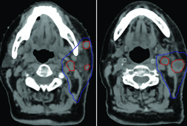
The first practical obstacle is that today’s patient is not the one on the previous planning CT. Weight loss, surgery, post-radiotherapy atrophy and a different setup shift organs at risk relative to the target. The second is that many patients do not return to the original department: the prior plan arrives as DICOM RT from another planning system, sometimes only as a printed isodose report. Combining those datasets requires image registration, and the choice between rigid and deformable is not neutral.
The Michigan protocol, described by Kelly Paradis and colleagues in Advances in Radiation Oncology in 2019, relied on rigid registration and acknowledged that this can introduce considerable uncertainty into composite dose metrics, to the point of making some organs impossible to assess. Deformable image registration (DIR) helps precisely in cases of extreme deformation, tissue loss and steep dose gradients, provided the deformation field is inspected. That is where QA tools such as grids and deformation heat maps come in. Radformation points to AutoContour, which pairs rigid and deformable registration with that kind of visualisation and more than 520 auto-segmentation models for recontouring organs at risk on the new CT. Recontouring with consistent nomenclature is what makes structures summable across datasets, the subject of TG-263 and of our target volume delineation guide.
Interval, tissue recovery and different fractionation: BED and EQD2
Adding the physical dose of two courses with different fractionation makes no radiobiological sense. A palliative 8 Gy single fraction and a curative 2 Gy-per-fraction schedule produce very different effects in late-responding tissues. The conversion runs through the linear-quadratic model and two everyday quantities:
$$BED = nd\left(1 + \frac{d}{\alpha/\beta}\right)$$
$$EQD_2 = D \cdot \frac{d + \alpha/\beta}{2 + \alpha/\beta}$$
Here n is the number of fractions, d the dose per fraction, D = nd the total dose and α/β the ratio describing how sensitive a tissue is to fractionation, roughly 2 to 3 Gy for late-responding tissues such as spinal cord and lung and about 10 Gy for most tumours. BED expresses the biologically effective dose; EQD2 translates any schedule into the equivalent dose delivered in 2 Gy fractions, which is what allows courses to be compared, summed and checked against published tolerance limits.
Then time enters. Michigan assigns each organ at risk dose discount factors stratified by the interval since the first treatment (0 to 3 months, 3 to 6 months and so on), with the DVH metric and the α/β fixed by institutional consensus. The factors are tissue-specific: the Radformation post notes that liver recovers far faster than lung. The Danish audit adds another layer: 61 distinct palliative and 104 curative prescriptions over the decade audited, from 8 Gy in a single fraction to 30 Gy in 10 fractions, which makes conversion to EQD2 mandatory rather than optional.
Plan sums and the conservative remaining dose
With images aligned and doses converted, the final step is the plan sum: overlaying the distributions and checking, organ by organ, how much of the cumulative limit has already been used. What is left is the remaining dose, which Michigan reports in physical dose at the fractionation planned for the new course so the dosimetrist has a usable number. When registration or α/β is uncertain, the prudent approach is the most conservative estimate: assume less recovery and the worst-case overlap. That is the reasoning ClearCheck, according to Radformation, automates in its latest release: recovery factors applied per organ, BED and EQD2 visualisation, a conservative remainder table for plan sums drawing on multiple registration sources, and the reference equations displayed alongside the result.
None of this replaces clinical judgement. ASTRO’s updated guidance on pancreatic radiotherapy illustrates how decisions hinge on site, interval and intent, and tolerance limits themselves are still being revised, as the ASTRO and VA DVH recommendations show. Predictive tools are entering the workflow too, from AI-predicted dose to automated plan checks.
What this means for medical physics practice
Most departments worldwide have neither a reirradiation registry nor a formal physics consult for retreatment. The cases exist and keep growing, yet they are often resolved by adding prescriptions by hand, exactly the practice the European consensus found in a quarter of the literature. The Michigan model can be replicated without heavy investment: a standard document listing α/β values, DVH metrics, interval-based discount factors and peer review, integrated with the existing TPS. Commercial registration and plan-check tools, such as those described by Radformation or the platforms in our auto-contouring software comparison, cut the manual work, but the department remains responsible for the parameters it feeds them.
The limitations are well known. The linear-quadratic model loses accuracy at high doses per fraction, results are sensitive to the chosen α/β, and rigid registration can make summation impossible where anatomy has deformed. The ESTRO/EORTC consensus asks for all of it to be documented, from registration method and α/β to interval and cumulative EQD2, and the Danish group is preparing a prospective registry to generate the data that are still missing. Until then the practical rule is simple: no reirradiation should begin without the full picture of what came before.
Source: Radformation Blog