
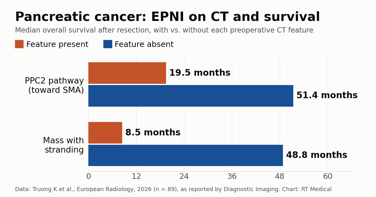
On preoperative CT of pancreatic cancer, the route and shape of tumor spread along nerves outside the gland may separate patients with very different outlooks. In a retrospective study of 89 patients who underwent resection for pancreatic ductal adenocarcinoma (PDAC), published in European Radiology, extra-pancreatic nerve plexus invasion (EPNI) along the PPC2 pathway, which leads to the superior mesenteric artery, and a “mass-with-stranding” morphology were both linked to markedly shorter overall survival. Median survival was 19.5 months with PPC2 involvement versus 51.4 months without it, and 8.5 months with mass-with-stranding versus 48.8 months without it.
The first author is Kathy Truong of the University of Miami Miller School of Medicine, with co-authors in Miami, Belgrade (Serbia) and Chicago. The paper was published online on October 22, 2026; its title refers to “early-stage” cancer because the associations held among stage I tumors.
How the 89-patient study was done
Of 265 “pancreatic tumor” cases in the institutional database from 2010 to 2020, 89 remained after excluding non-PDAC tumors (102), diffuse metastatic disease (73) and one duplicate. All had pathologically confirmed PDAC and pancreatic-protocol multidetector CT before resection; 54 were men, and the median age was 66 years.
Two experienced radiologists, blinded to outcomes, classified EPNI by pathway (PPC1, PPC2, anterior or mesenteric root) and by morphology (fine reticular, coarse reticular, nodular or mass with stranding). Survival was compared with stage-stratified log-rank testing and with Cox models adjusted for age, sex and surgical margin.
PPC2 and mass with stranding: the numbers
CT showed EPNI in 65 of 89 patients (73%), and its presence was associated with shorter overall survival (p < 0.001), including in stage I disease (p < 0.001). PPC2 involvement and mass-with-stranding morphology were each associated with worse survival (p < 0.001 for both) and stayed significant in stage I (p = 0.003 and p < 0.001). PPC2 spread was more common than PPC1 spread in stage II and stage III/IV disease than in stage I.
The abstract does not report hazard ratios or medians. According to Diagnostic Imaging’s account of the full text, multivariable analysis tied PPC2 involvement to a more than 2.5-fold higher risk of death and mass-with-stranding to a more than 2.6-fold higher risk. “The consistent association between PPC2 involvement on CT and adverse survival outcomes reinforces the role of PPC2 spread as a clinically meaningful imaging biomarker in PDAC,” the authors write.
What extra-pancreatic nerve plexus invasion is
Pancreatic adenocarcinoma has an unusual affinity for nerves. Perineural invasion is found in most resection specimens, and the tumor can track along nerve bundles out of the gland into the fat around the major arteries. These bundles form plexuses linking the pancreatic head to the celiac ganglia and to the nerve sheath of the superior mesenteric artery (SMA). The terminology dates to a 1958 surgical description by Yoshioka and Wakabayashi: plexus pancreaticus capitalis 1 and 2.
Put simply, PPC1 runs from the upper medial pancreatic head to the right celiac ganglion, near the celiac trunk. PPC2 starts at the uncinate process and follows the inferior pancreaticoduodenal artery to the SMA plexus. On CT, nerve invasion appears as soft-tissue strands, reticulation or nodules in the fat between the pancreas and the arteries; in the mass-with-stranding pattern, strands extend from the tumor toward the SMA. Inflammation, edema and fibrosis can mimic these findings, so the read depends on experience and good technique.
R0 margins, neoadjuvant therapy and NCCN criteria
The SMA margin is among the most frequently involved in surgery for pancreatic head cancer, and the artery is not routinely resected. One might assume PPC2 spread shortens survival simply by making an R0 resection harder. The study points elsewhere: both features predicted mortality independently of a positive margin. In the authors’ words, the imaging features “do not predict a technically difficult resection at the SMA margin, but may instead suggest aggressive tumor or microscopic infiltration.”
NCCN resectability criteria rest on solid tumor contact with the vessels: for pancreatic head tumors, SMA contact of up to 180° means borderline resectable disease, and more than that means locally advanced disease. The Society of Abdominal Radiology and American Pancreatic Association reporting template asks for stranding around the arteries to be described separately from solid contact, but the classification ignores the nerve pathway and the morphology of invasion. For resectable disease, NCCN already allows neoadjuvant therapy in the presence of high-risk features such as a markedly elevated CA 19-9, a large tumor or severe pain. The authors suggest that PPC2 and mass-with-stranding could help prioritize neoadjuvant chemotherapy, “although more studies are needed.”
The question also matters in radiation oncology. The region between the celiac trunk, SMA, portal/superior mesenteric vein and aorta, often called the “triangle,” concentrates perineural invasion and many local recurrences; its elective coverage is one of the topics in the updated ASTRO pancreatic cancer guideline, and the volumes in our guide to pancreatic cancer target delineation and RT planning already cover the celiac and SMA regions. As an editorial reading, a validated marker of PPC2 spread could help select patients for preoperative treatment. The study did not evaluate radiotherapy.
What changes for radiologists reading pancreatic CT
Nothing in the study requires new equipment. It used pancreatic-protocol CT with thin sections, pancreatic and portal venous phases, and multiplanar and maximum intensity projection (MIP) reconstructions. For departments in Brazil, Latin America and elsewhere, the immediate lesson is about technique and reporting: a routine abdominal CT makes fine strands around the SMA harder to see, and stating whether EPNI is present, along which pathway and with which pattern gives the tumor board more to work with.
Well-defined reading patterns also give automated tools a reference, like the models already tested for AI-based early pancreatic cancer detection. For patients who never reach surgery, MR-guided adaptive radiotherapy and ablative-dose radiotherapy are advancing.
Limitations: a small cohort and incomplete follow-up
The authors call the findings preliminary. Beyond the limits of a single-center retrospective design, they acknowledge the small cohort, loss to follow-up of about 20% of patients and a difference in median follow-up between the EPNI-positive and EPNI-negative groups. Splitting 65 cases across four pathways and four patterns also yields small subgroups and unstable risk estimates.
Only resected patients were included, and the 2010 to 2020 window spans shifts in neoadjuvant practice. The abstract does not report how many patients were treated before surgery or how well the readers agreed, which is key to reproducibility. External multicenter validation, correlation with nerve plexus pathology and trials testing whether acting on these findings changes outcomes are still needed.
Source: Diagnostic Imaging — Key Features with EPNI on CT May More than Double Mortality Risk in Pancreatic Cancer. Original paper: Truong K et al., European Radiology, 2026.