What is DICOM?
DICOM (Digital Imaging and Communications in Medicine) is an international standard for handling and storing medical images and associated information, such as patient information and treatment data. It is widely used in the healthcare industry to store, manage, and transmit medical images and information between different medical devices and systems.
DICOM plays an important role in keeping patient data safe by ensuring that medical images and information are stored and transmitted securely and consistently. DICOM images are stored in a standardized format, which ensures that images can be easily shared and viewed by different systems and healthcare professionals. DICOM also includes security features such as encryption and authentication, which protect patient data from unauthorized access and ensure that only authorized individuals can view and manipulate images.
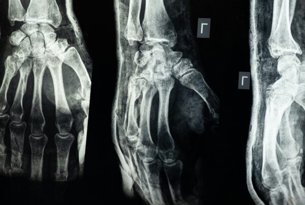
Medical Image Viewer
A DICOM viewer is a software application used to view and manipulate DICOM images. There are several features that a DICOM viewer must have, including:
- Image viewing and manipulation: The viewer must be able to display DICOM images in various modalities such as CT, MRI, and ultrasound, and must allow users to adjust image brightness and contrast, zoom in and out, and rotate images.
- Measurement tools: The viewer should have tools for measuring distances, angles, and areas within the images.
- Multi-frame support: The viewer must be able to display multi-frame DICOM images, such as those from a CT or MRI scan.
- Image annotation: The viewer should allow users to add annotations, such as text and shapes, to images for better communication and analysis.
- DICOM header information: The viewer must display DICOM header information, such as patient name, ID, study date, and modality.
- Compatibility with different systems: The viewer should be compatible with different DICOM-enabled systems, such as PACS (picture archiving and communication systems) and RIS (radiology information systems), to enable easy exchange of images and information.
- Security: The viewer should have security features such as user authentication and data encryption to protect patient data.
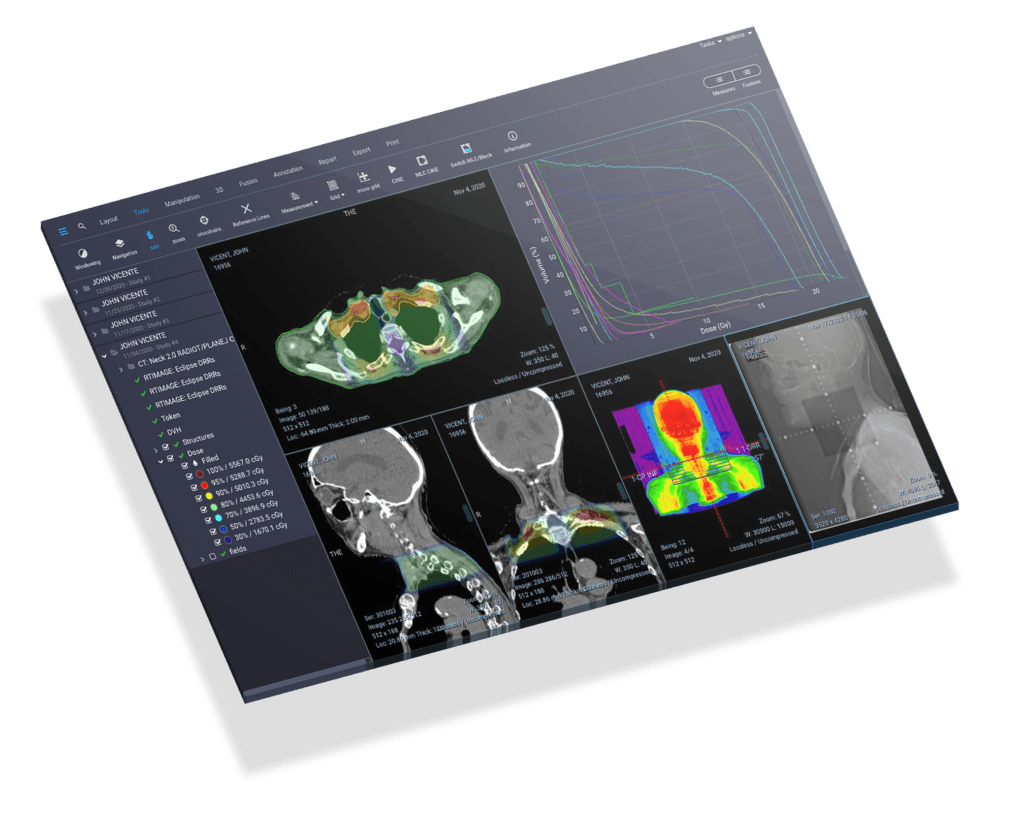
Radiotherapy Images
An image viewer used to view images of radiotherapy treatment plans should have certain tools to enable accurate and efficient evaluation of the treatment plans. Some of the key features that such a viewer should have include:
- Multi-modality support: The viewer should be able to display images from different modalities, such as CT, MRI, and PET, in order to provide a comprehensive view of the treatment area.
- 3D visualization: The viewer should be able to display 3D images of the treatment area, which allows for better visualization of the treatment volume and surrounding anatomy.
- Contouring tools: The viewer should have tools for contouring, which is the process of outlining the treatment area on the images. This allows the radiation oncologist to define the area that will be treated with radiation.
- Dose calculation and display: The viewer should be able to calculate and display the radiation dose distribution within the treatment area, which is essential for determining the appropriate treatment plan.
- Isodose lines: The viewer should be able to display isodose lines, which are lines that connect points of equal radiation dose on the image. This allows the radiation oncologist to visually assess the dose distribution and ensure that the desired dose is being delivered to the treatment area.
- Image fusion: The viewer should have the capability of fusing images from different modalities, such as CT and MRI, to provide a more accurate and detailed view of the treatment area.
- Multi-plan comparison: The viewer should be able to compare different treatment plans, which allows for more efficient and accurate evaluation of the plans.
- DICOM compatibility: The viewer should be able to read and display DICOM images, which is the standard format for storing and transmitting medical images and information.
- Security: The viewer should have security features such as user authentication and data encryption to protect patient data and ensure that only authorized individuals can view and manipulate the images.