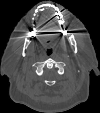
Metallic structures, such as metal implants or surgical clips, can pose a significant challenge for radiotherapists when it comes to outlining risk organs in tomography images. This is because metallic structures can cause artifacts in the images, which can make it difficult to accurately identify and outline the risk organs.
The main problem with metallic structures in tomography images is that they can cause streaks or shadows in the images, known as metal artifacts. These artifacts can obscure the underlying tissue and make it difficult to accurately identify and outline the risk organs. This can lead to errors in the treatment planning and deliver the wrong dose to the risk organs.
In addition to obscuring the underlying tissue, metal artifacts can also cause inaccurate dose calculations. This is because the metal absorbs some of the radiation, causing the dose to be higher than expected in areas close to the metal and lower than expected in areas farther away. This can lead to an increased risk of radiation-induced toxicity to healthy tissue and a decreased chance of the treatment being effective.
To overcome the problems caused by metallic structures in tomography images, radiotherapists often use specialized imaging techniques such as metal artifact reduction (MAR) or metal artifact reduction sequence (MARS) to reduce or eliminate the artifacts caused by metallic structures. These techniques can improve the visibility of the underlying tissue and allow for more accurate identification and outlining of the risk organs.
RT Medical Systems has developed a cloud-based software that aims to assist physicians in reducing metallic artifacts in CT images. The software utilizes advanced algorithms to eliminate the artifacts caused by metallic structures such as metal implants or surgical clips, which can make it difficult to accurately identify and outline risk organs in the images. By removing these artifacts, the software improves the visibility of the underlying tissue and allows for more accurate identification and outlining of the risk organs, which in turn can improve the accuracy of treatment planning and reduce the risk of radiation-induced toxicity to healthy tissue. The software is designed to be easy to use and integrate with existing systems, making it a convenient and efficient solution for physicians.
In conclusion, metallic structures can compromise the work of radiotherapists when it comes to outlining risk organs in tomography images by causing artifacts and errors in dose calculations. Specialized imaging techniques such as metal artifact reduction can help to reduce these problems and improve the accuracy of the treatment planning.


