
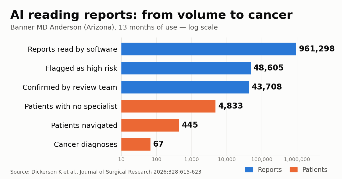
An artificial intelligence program that reads finalized radiology reports for high-risk incidental findings led to 67 cancer diagnoses over 13 months at Banner MD Anderson Cancer Center in Arizona. The study, published in the Journal of Surgical Research, reports 961,298 radiology reports screened by software, 43,708 findings confirmed by a dedicated team and 445 patients navigated to multidisciplinary care. It also shows that the narrowest point of the funnel was not reading the text but getting patients in front of a specialist.
The paper comes from Kylie Dickerson and colleagues at the Department of Surgery of the University of Arizona College of Medicine – Phoenix and the Department of Surgical Oncology at Banner MD Anderson. According to The Imaging Wire, the software is Azra AI, the company that in October announced it was merging with AI platform vendor Blackford to close the follow-up gap. The abstract itself does not name the vendor; it describes a commercial, end-to-end, oncology-focused natural language processing (NLP) solution. The authors report no disclosures and no funding.
How the Banner Health program worked
Banner Health is the largest integrated health system in Arizona, with 18 hospitals and more than 100 outpatient facilities, according to the paper. The software read reports from emergency department and outpatient imaging studies in real time and flagged those describing lesions at high risk for malignancy in the lung, liver and pancreas. The researchers supplied the keywords and parameters that defined a hit. Every flagged report went to a high-risk incidental findings team (HRIFT) for manual review. Patients with a new high-risk finding and no established oncologic care were then contacted and navigated to specialty care.
We checked the figures below against the PubMed abstract and the publicly visible sections of the article on ScienceDirect:
- 961,298 reports screened by the software;
- 48,605 (5.1%) flagged as containing a high-risk lesion;
- 43,708 confirmed by the team as clinically significant findings, which is 89.9% of flagged reports and 4.5% of all reports;
- 4,833 patients in the confirmed group without an established specialty provider, and therefore eligible for navigation;
- 445 patients successfully navigated to multidisciplinary care;
- 67 cancer diagnoses: 45 lung (67%), 14 liver (21%) and 8 pancreatic (12%).
In their conclusions, the authors write that the AI flagged a volume of lesions that “would have taken significantly more time to be worked up by traditional means or might have been missed altogether.”
Reading the percentages carefully
Some of the headline rates need unpacking. The “0.5% false-positive rate” reported in the study, and repeated in coverage, uses all screened reports as the denominator: 4,897 flagged-but-unconfirmed reports divided by 961,298. Measured against what the software actually flagged, roughly one alert in ten (10.1%) was rejected on human review. Seen from the other side, that is a positive predictive value of 89.9%. For anyone sizing a review team, that is the number that matters.
The 67 cancers equal 0.15% of validated reports, which the paper rounds to 0.2%, and 15% of navigated patients. The Imaging Wire wrote that Banner clinicians used the tool to review 961,300 reports; per the paper, the software read that volume and the human team reviewed the 48,605 flagged reports. The abstract also contains an inconsistency the available text does not resolve: it gives a study window of January 6, 2024 to June 30, 2025, roughly 18 months, yet presents the results as covering 13 months.
The figure missing from the outlet’s coverage may be the most useful one for practice. Of the 4,833 patients without a specialty provider, 445 (9.2%) were successfully navigated. The abstract does not say what happened to the rest: whether they declined, could not be reached, sought care elsewhere or were still in process.
Reading the report is not reading the image
Most commercial radiology AI analyzes pixels, looking for nodules, bleeds or fractures in the image itself. The Banner program does something different. It starts from the report the radiologist has already signed and searches the text for descriptions of suspicious lesions. It cannot find what the radiologist missed; it finds what was described and is at risk of never being acted on. That makes it a communication and logistics tool rather than a perception tool.
The weak link is well documented. The Fleischner Society recommendations for pulmonary nodules and the American College of Radiology (ACR) Incidental Findings Committee white papers on the liver, pancreas, kidneys and adrenals spell out when a finding warrants follow-up. No guideline ensures the recommendation reaches someone who will act on it, especially when the scan was done in the emergency department and the patient has been discharged, or when the ordering clinician is not the one who manages the patient. The authors cite a 2025 JAMA Internal Medicine study reporting missed diagnostic opportunities in 59% to 66% of lung and colorectal cancer patients who had primary care visits in the two years before diagnosis, with delays of 1 to 20 months. We have covered why lung nodule follow-up programs fail to scale and a pending lawsuit over an 8 mm nodule that went unfollowed and became stage 4 lung cancer, where the finding was in the report and the follow-up loop broke.
NLP on report text has its own pitfalls: negations (“no evidence of a mass”), hedged language (“cannot be excluded”), known lesions described as stable, and templates that repeat boilerplate phrases. Some of the 10% of rejected alerts likely reflects that kind of noise. Sensitivity, on the other hand, was not measured: unflagged reports were not audited, so nobody knows how many high-risk findings the software missed.
Lessons for imaging services and health systems
Three practical points come straight out of the numbers. First, the software cut nearly a million reports down to 48,600 for review, which still means about 3,700 reports a month for a human team. The cost of a program like this sits in staff time, not only in the license. Second, the final bottleneck was navigation rather than detection, which matches the experience of programs pairing AI with patient navigators in lung cancer screening. Third, performance depends on the keyword set and on how radiologists phrase their reports. Systems outside the US, including in Brazil and the rest of Latin America, would need to validate any such tool on reports written in Portuguese or Spanish before expecting similar yields.
Data governance is part of the picture. Scanning every report across a network to trigger outreach to patients requires a clear legal basis under health privacy rules, defined ownership of each alert and an audit trail. Products in the same space, such as Vital Guard, built to manage incidental radiology findings, and agentic AI tools that identify patients needing follow-up, run into the same question: who owns a finding once the report is signed?
Study limitations and what to watch
This is a single-system cohort with no control group and no comparison with the period before implementation. There is no way to tell how many of the 67 cancers would have been diagnosed anyway, or whether they were found at an earlier stage; the abstract reports neither stage nor time to diagnosis. The analysis is limited to lung, liver and pancreas, and the full text is paywalled, so this report relies on the abstract and the public sections of the article.
Even so, the paper provides something rare in this product category: peer-reviewed, system-wide numbers for a type of tool that has mostly been marketed with vendor-reported data. What would strengthen the case are before-and-after comparisons, time-to-treatment and stage-at-diagnosis measures, and audits of samples of unflagged reports to estimate sensitivity.
Source: The Imaging Wire — AI Finds Overlooked Cancers in Radiology Reports. Original paper: Dickerson K et al., Artificial Intelligence Guided Navigation of High-Risk Incidental Findings on Imaging Studies Leads to Cancer Diagnoses, Journal of Surgical Research, 2026.