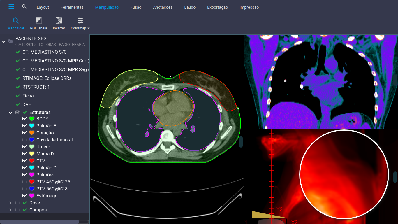
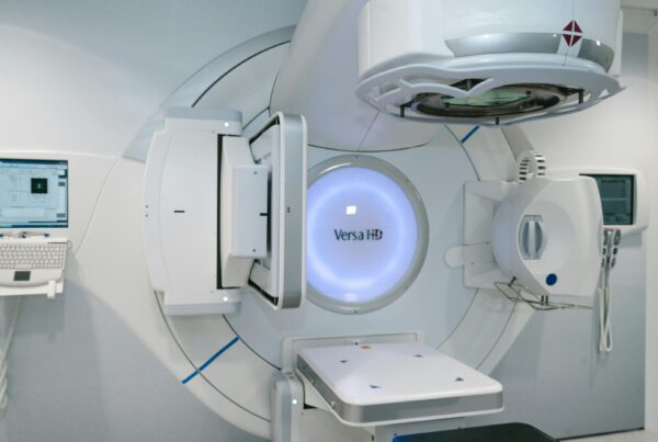
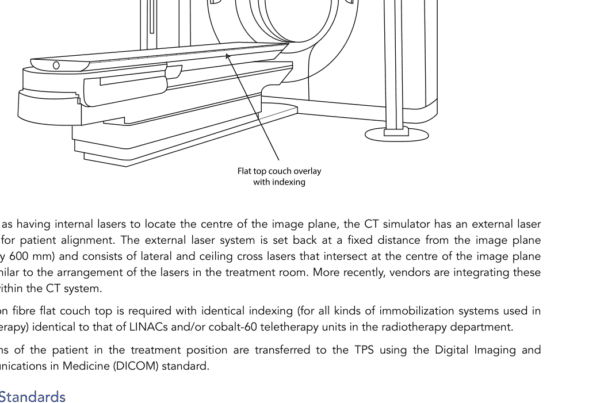
DICOM (Digital Imaging and Communications in Medicine) is an international communication standard that allows the transfer of digital medical information and images. The DICOM tags are numerical identifiers that describe specific information about an image or medical information.
Each image generated by medical equipment has stored inside a block of information about the technical aspects of the image, the patient and the transfer methods at the beginning, followed by the actual data of the image. Each DICOM tag has three components: the tag, the VR (Value Representation) and the name. The tag is a unique 16-bit identifier that identifies the information described in the tag. VR defines the type of information the tag represents, such as date, text, or number. The name is a textual description of the information described in the tag.
Some examples of DICOM tags include:
- (0010, 0010) Patient’s Name – the name of the patient described in the image or medical information
- (0010, 0020) Patient’s ID – the identification number of the patient depicted in the image or medical information
- (0020, 000D) Study Instance UID – a unique identifier for each medical study performed
DICOM tags guarantee security in the exchange of medical information, as they provide the unique and precise identification of each image or medical information, as well as providing specific information about the patient and the associated medical study. This allows healthcare professionals to identify and access the correct information for each patient, minimizing the possibility of medical errors.
DICOM file
The DICOM format uses a standardized file structure to store and transfer medical information, ensuring that information is preserved and interpreted consistently across different systems. This is critical to ensuring the integrity, security and privacy of medical information and protecting the patient. Formally known as ‘DICOM data objects’, they consist of various attributes (components), including a preamble (identifying the file type and components), a block of data (commonly known as DICOM headers, including patient demographic data, technical information about the image, the study, its acquisition parameters and acquisition device along with many other attributes listed) and the image data itself (a single attribute that contains the data needed to recreate the pixels or voxels of the image).
UIDs
Several unique identifiers (UIDs) are generated within each modality and included in the images
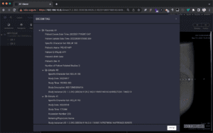
DICOM UIDs
produced. These identifiers together serve to present different information about the generating devices, the patient, the individual meeting and the files that make up the study. Moving on, each image within a study contains several different UIDs to link that single image to the rest of the series, the exam, and the overall patient encounter (the hierarchy being: Patient > Study > Series > Image). The UIDs generated as part of the process must be globally unique, and there are numerous issuance registries, which seek to avoid duplication by assigning batches to manufacturers and individuals or locations as needed.
All UIDs generated for DICOM services start with the first dig ¬1.2.840.10008[…] allowing for easy recognition among wider network traffic. Figure 8.2 identifies the UIDs in a portion of an example DICOM header.
Public tags and private tags
Public DICOM Tags
The public tags are the ‘common’ tags that have been standardized internationally by the committee and probably encountered under normal circumstances. These range from those common to all exams (patient name, date of birth, address, accession number, etc.) to those found only in certain exams (e.g. pitch, scan width, CT slice thickness ). Public tags have even group numbers (the first block of numbers on each line, such as [0008], [0010]).
Private DICOM Tags
On the other hand, private dicom tags found in medical image headers are distinguished from public tags because their group numbers are odd numbers.
Private tags contain pieces of image information that are unique to the equipment through which the image was acquired, or are extra pieces of data provided beyond what is available in public tags toto allow for more specialized use. Some uses of private tags can create a form of vendor lock-in – a problem seen historically in CT – without knowledge of the specific private tags and the expected data contained therein, it may only be possible to efficiently use a rebuild station of the same brand ( and maybe even model line) than the acquisition scanner.
Photometric interpretations
Not all digital images are captured in grayscale only; even when images are in grayscale, there is a question as to which ‘way’ the grayscale is applied to a particular image. For example, on a grayscale of 0-256, 0 is the whitest pixel value with 256 being pure black (a grayscale gradient in between) or vice versa.
The photometric interpretation definition is performed with each image and allows the display software to faithfully render (display) images as intended. The specialized literature states that problems were found with incorrect values of photometric interpretation, with images from legacy CR equipment being displayed inverted when transmitted by data sharing services until a patch for the original equipment was applied. The photometric interpretation values are typically:
Monochrome 2 (the smallest pixel value is displayed in black),
Monochrome 1 (the smallest pixel value is displayed in white) or RGB (color for display on ¬monitors). A photometric interpretation DICOM tag is included with each image to ensure images are displayed correctly.DICOM (Digital Imaging and Communications in Medicine) é um padrão internacional de comunicação que permite a transferência de informações e imagens médicas digitais. As tags DICOM são identificadores numéricos que descrevem informações específicas sobre uma imagem ou informação médica.
Cada imagem gerada por um equipamento médico possui armazenado em seu interior um bloco de informações sobre os aspectos técnicos da imagem, o paciente e os métodos de transferência em seu início, seguidos dos dados reais da imagem. Cada tag DICOM possui três componentes: a tag, o VR (Value Representation) e o nome. A tag é um identificador único de 16 bits que identifica a informação descrita na tag. O VR define o tipo de informação que a tag representa, como data, texto ou número. O nome é uma descrição textual da informação descrita na tag.
Alguns exemplos de tags DICOM incluem:
- (0010, 0010) Patient’s Name – o nome do paciente descrito na imagem ou informação médica
- (0010, 0020) Patient’s ID – o número de identificação do paciente descrito na imagem ou informação médica
- (0020, 000D) Study Instance UID – um identificador único para cada estudo médico realizado
As tags DICOM garantem a segurança na troca de informações médicas, pois proporcionam a identificação única e precisa de cada imagem ou informação médica, bem como fornecem informações específicas sobre o paciente e o estudo médico associado. Isso permite que os profissionais de saúde possam identificar e acessar as informações corretas para cada paciente, minimizando a possibilidade de erros médicos.
Arquivo DICOM
O formato DICOM utiliza uma estrutura de arquivo padronizada para armazenar e transferir informações médicas, garantindo que as informações sejam preservadas e interpretadas de maneira consistente em diferentes sistemas. Isso é fundamental para garantir a integridade, segurança e privacidade das informações médicas e proteger o paciente. Formalmente conhecidos como ‘objetos de dados DICOM’, eles consistem em vários atributos (componentes), incluindo um preâmbulo (identificando o tipo de arquivo e os componentes), um bloco de dados (comumente conhecido como cabeçalhos DICOM, incluindo dados demográficos do paciente, informações técnicas sobre a imagem, o estudo, seus parâmetros de aquisição e dispositivo de aquisição juntamente com muitos outros atributos listados) e os próprios dados da imagem (um único atributo que contém os dados necessários para recriar os pixels ou voxels da imagem).
UIDs
Vários identificadores exclusivos (UIDs) são gerados dentro de cada modalidade e incluídos nas imagens

UIDs DICOM
produzidas. Esses identificadores juntos servem para apresentar diferentes informações sobre os dispositivos geradores, o paciente, o encontro individual e os arquivos que compõem o estudo. Continuando, cada imagem dentro de um estudo contém vários UIDs diferentes para vincular essa única imagem ao restante da série, ao exame e ao encontro geral do paciente (sendo a hierarquia: Paciente > Estudo > Série > Imagem). Os UIDs gerados como parte do processo devem ser globalmente exclusivos e existem vários registros de emissão, que procuram evitar a duplicação, atribuindo lotes a fabricantes e indivíduos ou locais conforme necessário.
Todos os UIDs gerados para serviços DICOM começam com o primeiro dig ¬1.2.840.10008[…] permitindo seu fácil reconhecimento entre o tráfego de rede mais amplo. A Figura 8.2 identifica os UIDs em uma parte de um exemplo de cabeçalho DICOM.
Tags públicas e tags privadas
Tags DICOM Públicas
As tags públicas são as tags ‘comuns’ que foram padronizadas internacionalmente pelo comitê e provavelmente encontradas em circunstâncias normais. Estes vão desde os comuns em todos os exames (nome do paciente, data de nascimento, endereço, número de acesso, etc.), até aqueles encontrados apenas em determinados exames (por exemplo, pitch, largura da varredura, espessura do corte na TC). As tags públicas têm números de grupos pares (o primeiro bloco de números em cada linha, como [0008], [0010]).
Tags DICOM Privadas
Já as tags dicom privadas encontradas em cabeçalhos de imagens médicas são diferenciadas de marcas públicas por seus números de grupo serem números ímpares.
As tags privadas contêm partes de informações de imagem que são exclusivas do equipamento por meio do qual a imagem foi adquirida ou são partes extras de dados fornecidas além daquelas disponíveis em tags públicas para permitir um uso mais especializado. Alguns usos de tags privadas podem criar uma forma de aprisionamento do fornecedor – um problema visto historicamente em CT – sem o conhecimento das tags privadas específicas e dos dados esperados contidos nelas, só pode ser possível usar com eficiência uma estação de reconstrução da mesma marca (e talvez até mesmo linha de modelo) que o tomógrafo de aquisição.
Interpretações fotométricas
Nem todas as imagens digitais são capturadas apenas em escala de cinza; mesmo quando as imagens estão em escala de cinza, há uma dúvida sobre qual ‘maneira’ a escala de cinza é aplicada em uma imagem específica. Por exemplo, em uma escala de cinza de 0-256, 0 é o valor de pixel mais branco com 256 sendo preto puro (um gradiente de tons de cinza entre) ou vice-versa.
A definição da interpretação fotométrica é realizada com cada imagem e permite que o software de exibição renderize (exiba) imagens fielmente conforme pretendido. A literatura especializada afirma que foram encontrados problemas com valores incorretos de interpretação fotométrica, com imagens de equipamentos CR herdados sendo exibidas invertidas quando transmitidas por serviços de compartilhamento de dados até que um patch para o equipamento original fosse aplicado. Os valores de interpretação fotométrica são tipicamente:
Monocromático 2 (o menor valor de pixel é exibido em preto),
Monocromático 1 (o menor valor de pixel é exibido em branco) ou RGB (cor para exibição em ¬monitores). Uma tag DICOM de interpretação fotométrica está incluída em cada imagem para garantir que as imagens sejam exibidas corretamente.

