

Choroid Plexus Shows Detectable MRI Changes in Long COVID Patients
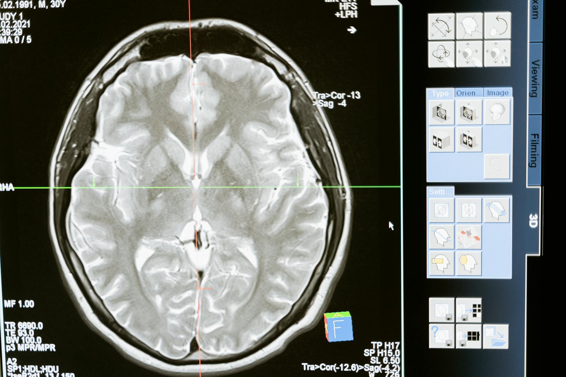
Researchers at NYU Langone Health have identified, through advanced magnetic resonance imaging, that patients with Long COVID exhibit measurable abnormalities in the choroid plexus — a brain structure responsible for cerebrospinal fluid production and immune signaling. Published in Alzheimer’s & Dementia, the finding establishes a concerning link between persistent COVID-19 effects and Alzheimer’s-associated biomarkers.
The study evaluated 179 participants divided into three groups: 86 Long COVID patients, 67 who fully recovered from COVID-19, and 26 COVID-naive controls. Results reveal significant structural and vascular differences that may carry profound implications for neurology.
Key MRI Findings
Imaging demonstrated that choroid plexus volume in Long COVID patients was 10% larger compared to fully recovered individuals. Beyond the volumetric increase, reduced blood flow through choroid plexus vessels was observed, indicating vascular dysfunction.
What makes this finding particularly relevant is its correlation with Alzheimer’s-associated biomarkers. Choroid plexus changes correlated with elevated levels of pTau217 and GFAP — proteins that are known markers of neurodegeneration. Clinically, patients showed cognitive decline of approximately 2% on the Mini-Mental State Exam. This underscores the importance of advanced imaging tools, something the evolution of PACS technology and AI has been driving forward.
Mechanism: Inflammatory Vascular Remodeling
According to the researchers, persistent inflammatory reactions following COVID-19 infection may trigger a process of “vascular remodeling” in the choroid plexus. This process involves thickening and scarring of the vascular lining, restricting blood perfusion and potentially compromising cerebrospinal fluid production and blood-CSF barrier integrity.
Senior author Dr. Yulin Ge stated: “Physical, molecular, and clinical evidence suggests that a larger choroid plexus may be an early warning sign of Alzheimer’s-like cognitive decline.” This observation positions MRI as a potentially crucial tool for early screening of post-COVID neurodegeneration.
Implications for Radiological Practice
For radiologists, this study reinforces the importance of brain MRI protocols that include volumetric and perfusion assessment of the choroid plexus, especially in patients with Long COVID history and cognitive complaints. The ability to detect these changes early could pave the way for preventive interventions.
Healthcare PACS systems improving telemedicine will need to support efficient visualization and quantification of these metrics, reinforcing the need for integration between post-processing tools and diagnostic workstations.
Next Steps in Research
The research team plans longitudinal follow-up studies to determine whether identified brain changes predict future cognitive decline development. This approach could define whether choroid plexus MRI should become part of post-COVID follow-up protocols.
For the radiology community, these findings represent yet another example of how advanced imaging can reveal subtle pathological processes before they manifest clinically, reinforcing the diagnostic and predictive role of the specialty.
Source: Applied Radiology