

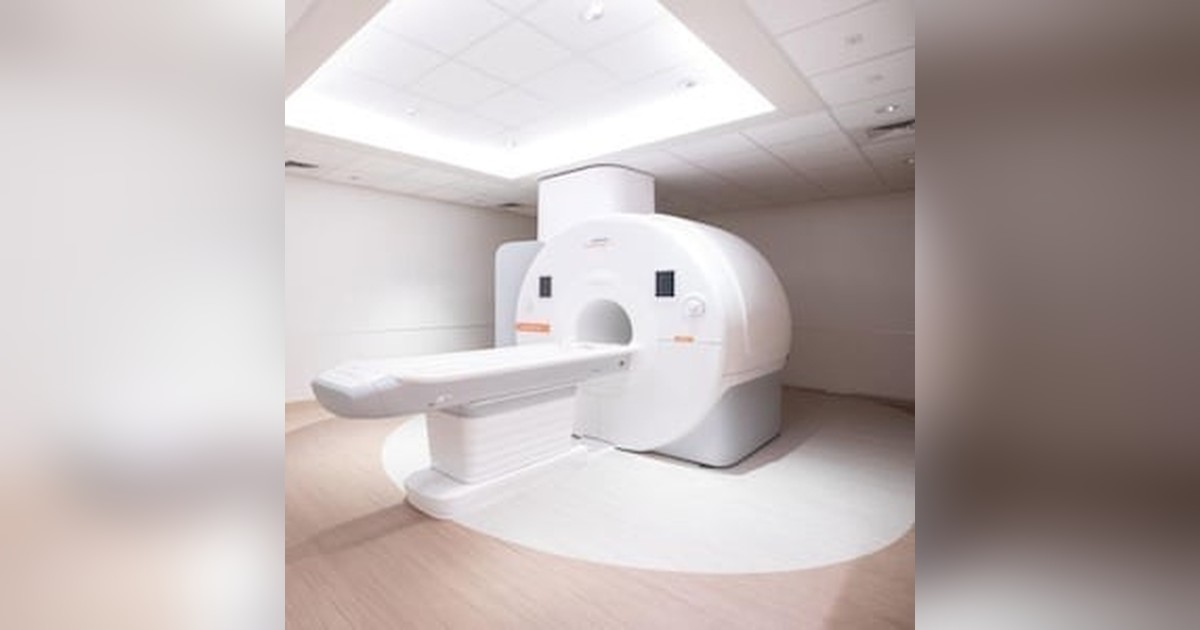
Yale New Haven Hospital has installed a PET/MRI system and will become the first facility in Connecticut to offer the hybrid exam, which combines positron emission tomography and magnetic resonance imaging in a single session. The roughly 9-ton scanner will sit inside the Adams Neurosciences Center, the hospital’s new neurology and neurosurgery complex, scheduled to open in 2027. The announcement, dated September 29 and published by ITN on October 5, points to applications in Alzheimer’s disease, epilepsy and oncology.
What Yale New Haven announced
According to the release, the new system records metabolic and molecular information (from PET) and anatomical information (from MRI) within the same exam. The hospital notes that it also opened Connecticut’s first MRI center, in 1986. Neither the release nor the ITN report names the manufacturer, model or investment. Minutes from an October 2024 certificate-of-need hearing at Connecticut’s Office of Health Strategy only state that the neurosciences tower project includes one PET-MR and one intraoperative MRI system, without identifying a vendor.
Christopher Whitlow, MD, PhD, radiologist-in-chief at the hospital and chair of radiology at Yale School of Medicine, framed the rationale simply: performing both exams at once lets clinicians see structure and function together, giving a fuller picture of the patient’s condition. He said the scanner will matter for both patient care and research, particularly in neuroscience and oncologic imaging.
The Adams Neurosciences Center will span more than 500,000 square feet across two patient towers. Planned services include epilepsy, movement disorders such as Parkinson’s disease, spine disorders, stroke and aneurysm care, and neuromodulation therapies for pain. The building will also house dedicated neurosurgery operating rooms, rehabilitation, all-private inpatient rooms and research space.
How simultaneous PET/MRI works
Putting PET and MRI in one gantry required solving an engineering problem: conventional photomultiplier tubes in PET detectors do not work inside a strong magnetic field. Today’s integrated systems use solid-state photodetectors, such as avalanche photodiodes or silicon photomultipliers (SiPM), which tolerate the field and, in the SiPM case, support time-of-flight (TOF) measurement. The PET ring sits inside the magnet, so both acquisitions happen at the same time rather than sequentially as in PET/CT.
Simultaneity brings its own advantages. Spatial registration between the two datasets is intrinsic, and MR sequences can be used to correct patient motion in the PET data. It also becomes possible to study fast-changing processes, for instance comparing MR perfusion with tracer uptake over the same time window.
The hardest part is attenuation correction. In PET/CT, the CT itself provides a tissue density map that is converted into attenuation coefficients at 511 keV. MRI does not measure electron density, so that map must be estimated. Early solutions segmented Dixon images into air, lung, fat and soft tissue while ignoring bone, which tends to underestimate uptake near bony structures. Atlas methods followed, then ultrashort or zero echo time (UTE/ZTE) sequences that depict cortical bone, and more recently deep learning methods that synthesize a “pseudo-CT” from MR data. In the brain, these techniques have substantially improved quantification, a requirement for dementia studies.
Where the exam makes a difference
Yale’s release lists the modality’s classic uses. In drug-resistant epilepsy, FDG PET can reveal hypometabolism in the epileptogenic zone while high-resolution MRI looks for cortical dysplasia or hippocampal sclerosis; fusing both helps localize the focus before surgery, including in patients whose initial MRI is negative. In dementia, combining FDG, amyloid or tau PET with MR volumetry and structural sequences lets a team assess neurodegeneration patterns and atrophy in one appointment, a topic that connects with recent work on MRI-measured brain aging.
In oncology, PET/MRI fits where MRI is already the reference for soft tissue: pelvis, liver, head and neck, central nervous system and spine. Prostate cancer is a good example, and PSMA PET combined with multiparametric MRI is already under regulatory review, as shown by the FDA Fast Track designation for pre-biopsy PSMA PET plus MRI. In pediatrics, dropping the CT component reduces ionizing radiation exposure, which matters for children with lymphoma or sarcoma who undergo repeated follow-up scans. The tracer dose remains, but the CT dose disappears.
There are known limitations. Detection of small lung nodules is inferior to CT, protocols tend to be longer, and patients must pass MR safety screening, which rules out or complicates some implants. For anyone tracking nuclear medicine, as in the discussions at SNMMI 2026, PET/MRI is increasingly tied to new radiotracers and theranostics.
Cost, adoption and the Latin American context
More than a decade after the first commercial simultaneous systems arrived, PET/MRI remains a niche technology. The scanner is expensive, installation requires MR shielding plus radiopharmacy infrastructure, and staff must master both modalities. Reimbursement also weighs on adoption: many health systems have no dedicated payment for the hybrid exam, which ends up billed as the sum (or part) of separate procedures. As a result, the modality tends to cluster in academic centers with research programs, a profile shared by Yale and by other institutions that sign long-term partnerships with imaging vendors.
In Brazil and across Latin America, PET/MRI units are few and concentrated in large private and research centers. For departments considering a purchase, the U.S. experience suggests starting with indications where the gain is clearest, such as epilepsy, neuro-oncology, pediatrics and pelvic imaging, and planning early for PACS integration and joint reporting between radiology and nuclear medicine. In radiation oncology, fusing PET and MRI acquired in the same position is also attractive for target delineation, provided geometry and coils are compatible with treatment planning.
What to watch next
Yale’s announcement is about installation, not clinical results: the scanner will not see patients until 2027. It remains to be seen what exam volume the service will reach, which protocols it will prioritize and how scanner time will be split between clinical care and research. It is also worth watching whether the hospital discloses the manufacturer and system configuration, which the release omits. For radiology, the case reinforces a trend: dedicated neuroscience centers are building molecular imaging into routine care rather than treating it only as a research tool.
Source: ITN (Imaging Technology News)



