
A one-minute scan beat the standard-of-care exam
A long-axial-field-of-view PET/CT produced diagnostic-quality images from just one minute of
acquisition — and even at that extreme the readers scored it above the conventional PET/CT used in
daily practice. The result was presented on October 18 at the annual meeting of the European
Association of Nuclear Medicine (EANM) in Vienna by Cameron Pain, of the Peter MacCallum Cancer
Centre in Melbourne, Australia.
“A one-minute scan on this system gave us images that our readers rated more highly than the
standard-of-care scans we perform every day,” Pain said in an EANM news release. “That was the
striking finding: even when we cut the acquisition time by 90%, image quality exceeded current
standard-of-care methods.”
How the study was built
Twenty-four patients scheduled for oncological PET/CT took part, each receiving one of five
radiotracers. Every participant had a standard-of-care scan on a conventional short-axial-field-of-view
(SA-FOV) system and, immediately afterwards, a 10-minute acquisition on the long-axial-field-of-view
(LA-FOV) Omni Legend 128 from GE HealthCare — a scanner with a 128 cm axial field of view that
covers essentially the whole body in a single bed position.
From the raw data of that single 10-minute acquisition, the researchers reconstructed images
equivalent to 5, 3, 2 and 1 minute of counting time. Two nuclear medicine specialists, blinded to
acquisition time, graded each set on a five-point scale for overall diagnostic quality, sharpness,
lesion conspicuity (how clearly a lesion stands out from surrounding tissue) and image noise.
The numbers behind the headline
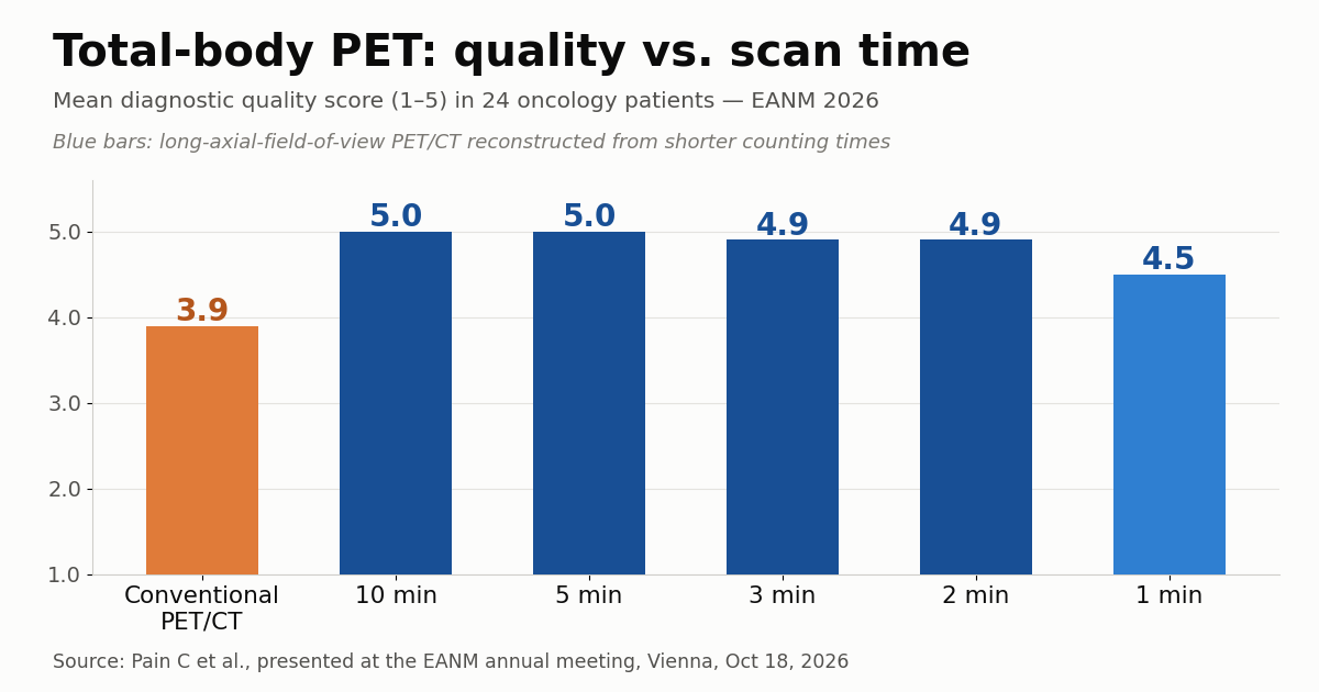
Mean diagnostic quality scores were 5.0 for the 10- and 5-minute reconstructions, 4.9 for the
3- and 2-minute reconstructions and 4.5 for the 1-minute set. Conventional PET/CT scored 3.9 and
posted lower averages than the LA-FOV system across every assessed category and every acquisition
time. Put plainly: the most aggressive reconstruction on the new scanner still read better than a
full-length exam on the old one.
It is worth saying why that is physically plausible. A PET scanner detects annihilation photon
pairs in coincidence; the longer the axial field, the larger the solid angle covered and the greater
the fraction of emitted events that actually reaches the detectors. A 128 cm system captures roughly
an order of magnitude more counts per unit time than a 20 to 25 cm one. Because signal-to-noise in
PET scales approximately with $\sqrt{N}$, where $N$ is the number of acquired counts, that
sensitivity gain can be spent on time, on dose or on image quality — which is exactly the author’s
argument.
Three currencies, one sensitivity gain
“Total-body PET gives us choices we haven’t had before. We can use its sensitivity to scan
patients faster, to lower the radiation dose and to see disease more clearly,” Pain said. That is
the right reading: the gain is singular and has to be allocated.
Shorter scans affect comfort directly, especially for patients in pain or unable to lie still,
and they cut motion blurring — a problem we examined when we looked at
the cost of motion-related repeat sequences in MRI.
They also raise daily throughput and, through higher sensitivity, allow less radiotracer per
patient, which matters economically in departments that depend on short-half-life
radiopharmaceuticals.
What it means for practice and for the installed base
No department replaces a PET/CT on the strength of a conference abstract, but the replacement
conversation shifts when the argument stops being “nicer images” and becomes installed capacity. If
one scanner does in 2 minutes what another does in 20, the return calculation no longer hinges on
purchase price alone: throughput per shift, tracer consumption and the ability to scan patients who
cannot tolerate a long protocol all enter the equation.
There is a rarely mentioned counterweight. Very short protocols increase departmental load in
everything that is not acquisition — preparation, venous access, uptake time, positioning and
reporting — and those steps do not shrink proportionally. The bottleneck tends to migrate from the
scanner room to the schedule. LA-FOV systems also generate larger data volumes per study, with
direct consequences for storage and network, a point we raised when analysing
high-cost hybrid scanner installations.
The limits of what was shown
The author himself drew the boundaries. This is 24 patients, with a subjective reading endpoint
rather than diagnostic accuracy measured against a reference standard. Larger studies will need to
confirm that very short acquisitions do not miss small or faint lesions — the scenario where
reduced counts should hurt most. And it remains to be seen how fast scanning behaves in real
workflow: scheduling, tracer logistics and patient experience do not show up in an image quality
score.
Even so, the direction is consistent with what other groups have reported since the first
long-axial-field systems arrived. The debate is no longer whether the extra sensitivity exists, but
how to spend it — on speed, on dose or on detection. For anyone following
the growing use of PET in targeted diagnostic pathways,
it is a choice that will keep surfacing in protocol design.
Source: AuntMinnie — EANM: Next-generation PET scanner cuts imaging time to one minute