
Handheld screening ultrasound of the breast has predictable blind spots: cancers in the left breast, in peripheral or fatty areas, and in the upper inner quadrant are the most likely to go undetected. That is the message of a Yale School of Medicine study to be presented at RSNA 2026 on December 3, which compared cancers found and missed on screening whole-breast ultrasound (WBUS).
The study, led by Katie Jolin, MD, reviewed 32,116 digital breast tomosynthesis (DBT) screenings performed between 2020 and 2023, about 64% of which included same-day WBUS. The goal was to pin down which features let a cancer slip past the technologist-performed handheld exam and surface only later, at targeted diagnostic workup.
What the Yale team measured
The researchers compared cancers not detected on screening WBUS but found at subsequent diagnostic workup with those the exam caught. Three radiologists reviewed each tumor for size, subtype, laterality, quadrant, and surrounding tissue composition. In all, 141 cancers were diagnosed across the exams; among the women screened with ultrasound, 79 were detected by WBUS and 20 were initially missed. About 90% of the tumors were invasive in both groups.
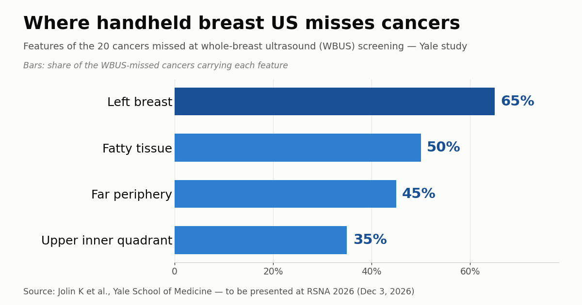
Detected cancers were larger than missed ones (mean 13.6 mm vs. 9.1 mm), which helps explain why small lesions are harder to catch at screening. Laterality stood out too: detected cancers split almost evenly between the right (49%) and left (51%) breasts, while 65% of missed cancers were in the left breast.
The upper inner quadrant was the most common site for missed cancers (35%), whereas detected cancers clustered in the upper outer quadrant (40%). Tissue composition drew the sharpest contrast: 88% of detected cancers lay within fibroglandular tissue, while half (50%) of the missed cancers were in fatty tissue (p < 0.01). Nearly half of the missed tumors (45%) were in the far periphery of the breast.
Why handheld ultrasound lets some tumors slip
Ultrasound is a supplemental screening option for women with dense breasts, where fibroglandular tissue both hides lesions on mammography and raises risk. It adds cancer detection beyond mammography and DBT, but it is operator dependent: in handheld WBUS a technologist sweeps the probe across the breast in real time, and coverage and sensitivity vary with technique and experience. Tellingly, some cancers invisible at screening are easily seen afterward, once the diagnostic exam is aimed at the finding.
The results have a physical logic. In fatty tissue a hypoechoic mass contrasts less with its surroundings and blends into the fat, unlike in fibroglandular tissue; and the far periphery, near the chest wall and the margins, is harder to cover completely in a handheld sweep. That performance gap helps explain why glandular tissue on breast ultrasound behaves so differently from fatty tissue when it comes to flagging a tumor.
This is also where automated breast ultrasound (ABUS) enters the conversation. Instead of a manual sweep, ABUS acquires standardized breast volumes that the radiologist reviews afterward; that lowers operator dependence but trades the dynamic, real-time exam for an offline read. Neither approach erases blind spots — it shifts them — and the Yale study maps exactly where handheld WBUS tends to fall short.
What it changes in practice
In Brazil and much of Latin America, ultrasound is a mainstay of supplemental screening in dense breasts because it is inexpensive, widely available, and does not depend on limited MRI capacity. Since handheld WBUS dominates over ABUS in public and private services, performance rests on the scanning protocol and the training of whoever runs it. Knowing that the left breast, the periphery, and fatty regions concentrate the failures is a practical argument to standardize coverage and sharpen attention to those territories.
The takeaway is not to abandon the method but to calibrate expectations. Ultrasound still adds cancers that mammography does not show — the same debate raised when asking whether screening ultrasound is still needed in the DBT era. The study reinforces that breast density, laterality, and location belong in how we read the risk of a negative exam — and that a WBUS with no findings does not close the case when a symptom or clinical change is present.
Limitations and what to watch
This is a single-center, retrospective study to be presented at RSNA 2026 that has not yet been peer reviewed or published in a journal. The number of missed cancers is small (20 cases), so figures such as the 35% for the upper inner quadrant rest on a small denominator and may shift. The definition of “missed” — not detected at WBUS screening but found at later workup — also depends on how and when follow-up happened.
Even so, the map of blind spots is useful and converges with other recent work gathered in the roundup of the breast imaging studies that defined 2026. Watch for the final data when the talk takes place on December 3, and whether the pattern holds at other institutions, with different scanning protocols and with ABUS. For now, the message is concrete: small cancers, in the left breast, in fatty or peripheral areas, and in the upper inner quadrant deserve extra attention on handheld screening ultrasound.
Source: AuntMinnie — Handheld screening ultrasound has blind spots