MRI read with the O-RADS system identified endometriosis-associated ovarian cancer with 86.8% accuracy in a study to be presented at RSNA 2026, offering a more objective way to decide which indeterminate adnexal masses need oncologic surgery. The work, carried out at the University of Montreal, focused on patients whose ultrasound had already rated the lesion as intermediate- or high-risk — exactly the setting where endometriosis most confounds assessment.
The results will be shown in an RSNA 2026 session on December 1 and have not yet appeared in a peer-reviewed journal. Even so, the figures stand out for their high sensitivity to malignant tumors in a notoriously difficult patient group.
What the University of Montreal study measured
The team led by Catherine Wang, MD, studied 143 women with endometriosis who underwent pelvic MRI for an indeterminate adnexal mass — lesions rated O-RADS 3, 4, or 5 on ultrasound. Every patient went on to surgery with pathological analysis, which let the researchers compare the imaging score against the true diagnosis. Two radiologists independently assessed morphological and functional MRI features and assigned O-RADS MRI scores.
The final cohort comprised 143 patients, with a median age of 45 years, and 182 adnexal masses. On pathology, 65.9% of the lesions were benign, 14.8% were borderline tumors and 19.2% were invasive tumors. Among the borderline tumors, 48.1% were of the seromucinous subtype; among the invasive tumors, 62.8% were clear-cell or endometrioid carcinomas — the very histologies most tied to endometriosis.
Which imaging findings pointed to malignancy
Seven factors were linked to malignancy, all with p < 0.01: solid tissue, size over 4.5 cm, multilocularity, loss of T2 shading, an abnormal ovarian pedicle, menopausal status and deep pelvic endometriosis. Loss of T2 shading matters in particular because that sign typically characterizes a classic endometrioma; its absence can signal transformation.
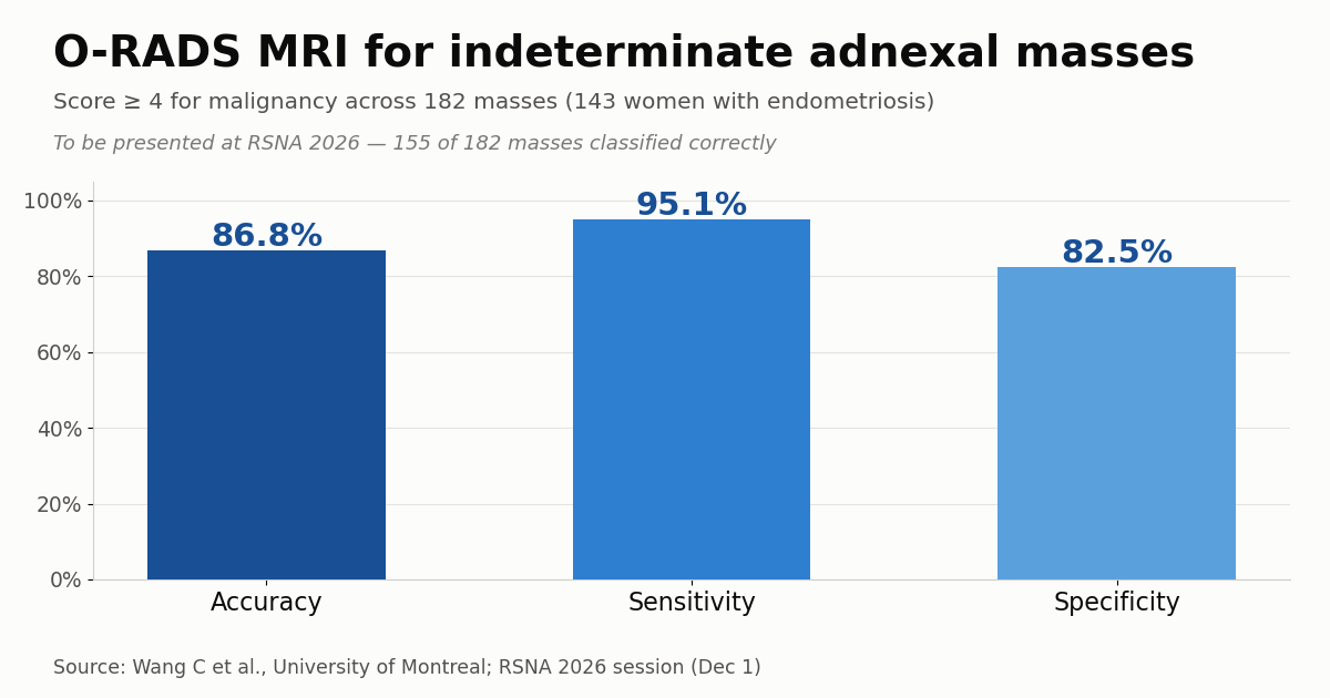
An O-RADS MRI score of 4 or higher correctly classified 155 of the 182 masses, for an accuracy of 86.8%, sensitivity of 95.1% and specificity of 82.5%. Invasive tumors differed from borderline ones by the look of the solid tissue: obtuse angles (odds ratio, OR, 5.8), irregular borders (OR 4.0) and fewer than four solid nodules (OR 7.0). Beyond that, every tumor with an apparent diffusion coefficient (ADC) of 0.7 or lower (× 10⁻³ mm²/s) in the solid tissue proved to be invasive, an almost dichotomous threshold in this sample.
Why endometriosis complicates reading adnexal masses
O-RADS MRI (Ovarian-Adnexal Reporting and Data System) is a structured American College of Radiology scheme that scores adnexal masses from 1 to 5 by their probability of malignancy, combining morphology with the dynamic contrast-enhancement pattern. It was designed above all to resolve the lesions ultrasound leaves indeterminate, guiding follow-up or surgery more confidently. It follows the same logic as imaging scores already used elsewhere, such as the breast MRI score for neoadjuvant response and MRI scores for renal masses.
Endometriosis makes the task thornier for two reasons. First, endometriomas and other atypical benign lesions can mimic neoplasms on imaging. Second, the disease raises the risk of specific ovarian cancers — chiefly clear-cell and endometrioid carcinomas — and of seromucinous borderline tumors. That excess risk is histology-specific and modest in absolute terms, but it reshapes how a suspicious mass should be weighed in a patient who already has the disease. The upshot is a large number of masses that read as indeterminate on ultrasound, precisely the ones the study set out to reclassify with MRI, where diffusion-weighted sequences and the ADC add functional information that grayscale ultrasound cannot.
What changes in triage and surgical referral
In practice, an objective score with high sensitivity helps separate the patients who need oncologic surgery from those who can follow conservative management or a less aggressive operation. According to the researchers, MRI supports more consistent reporting and appropriate surgical referral while helping to avoid unnecessary aggressive interventions. Imaging-based stratification dovetails with other decision tools in gynecologic oncology, much as MRI has reshaped prostate cancer staging.
For health systems where endometriosis is common and access to pelvic MRI and standardized reporting is uneven — including much of Latin America — a reproducible protocol like O-RADS MRI can harmonize decisions and cut reoperations. Where MRI capacity already exists, folding the score into the adnexal-mass work-up should sharpen referral to the gynecologic oncologist rather than add imaging for its own sake.
Limitations and what to watch
Because this is a single-center analysis with a surgical endpoint, the cohort is already enriched for suspicious lesions, which inflates the prevalence of malignancy and may not reflect the general population. The 82.5% specificity means some benign masses still receive a high score, with a risk of unnecessary surgery. As research to be presented at a meeting, the data still need peer-reviewed publication and external validation before they change routine practice. The double read by two radiologists also leaves the score’s reproducibility outside specialized centers an open question to track once the full study appears.
Source: AuntMinnie — O-RADS MRI spots ovarian cancer in women with endometriosis