

Ninety minutes of training to work up DVT at the bedside
The FDA has granted 510(k) clearance to ThinkSono Guidance, the first AI-powered ultrasound guidance software cleared in the United States for the evaluation of suspected lower-extremity deep vein thrombosis. The number that matters more than the “first” label: healthcare staff with no ultrasound training captured diagnostic-quality images in 87.1% of cases after 60 to 90 minutes of standardized training.
What exactly was cleared
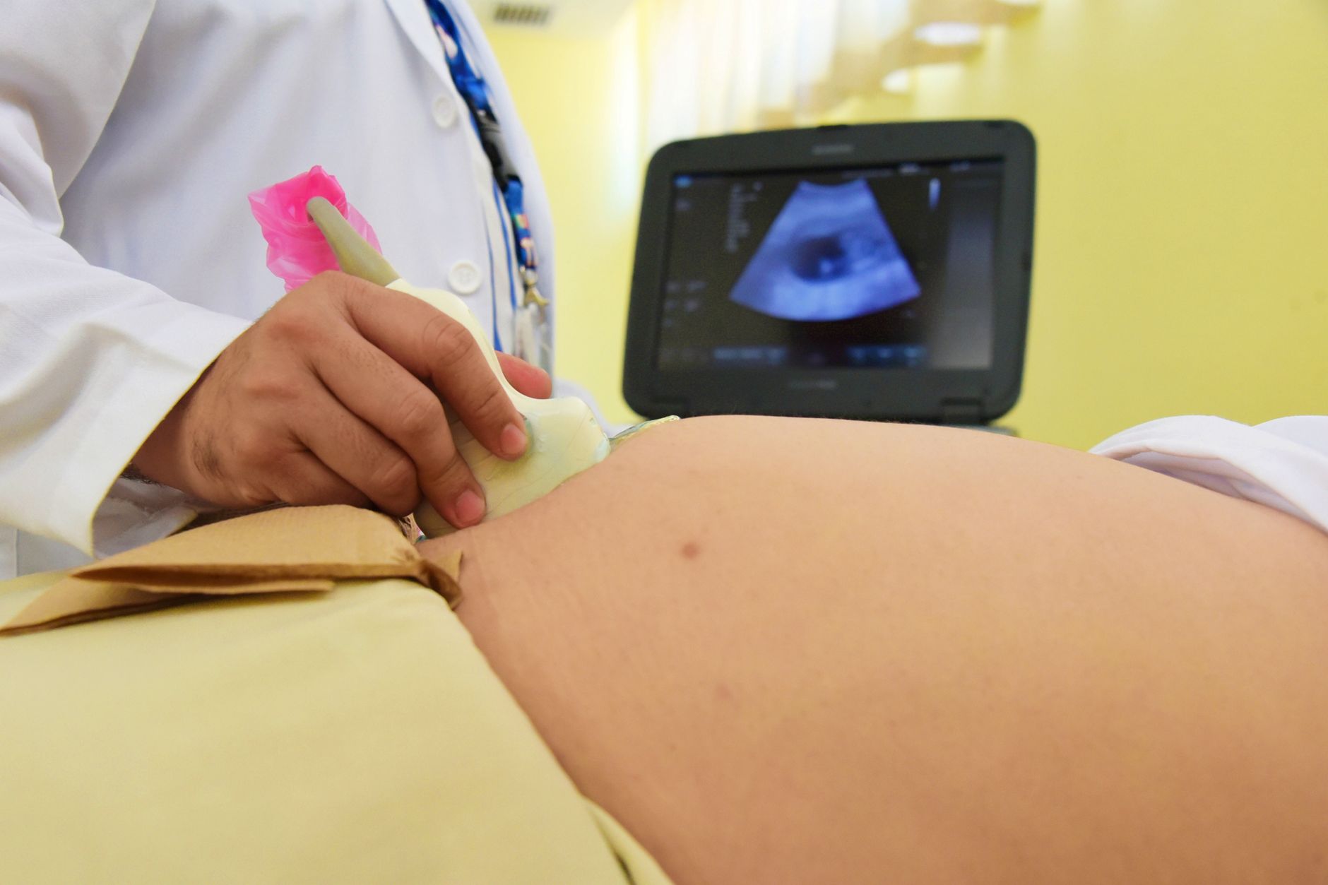
The product is software, not a scanner. It runs as a mobile app paired with compatible Clarius handheld point-of-care ultrasound probes and walks the operator through acquisition: where to place the probe, how much pressure to apply during the compression maneuver, and how to hold anatomical alignment along the proximal deep venous system. The device had received FDA Breakthrough Device Designation in February 2026, which accelerated the regulatory pathway.
The boundary is worth underlining: the algorithm guides acquisition. It addresses the shortage of trained hands at the point of care, not interpretation, which remains a physician responsibility over the compression cine-loops the app produces.
The validation numbers
The submission rests on a double-blinded, multicenter study of 1,691 patients across 24 U.S. and international sites. With non-expert operators using the software:
- 87.1% of exams reached adequate diagnostic image quality;
- 92.9% sensitivity for DVT;
- 97.1% specificity.
A double-blinded design at that sample size is unusual for imaging software — most clearances of this kind rest on retrospective cohorts of a few hundred exams. And in this particular use case, 97.1% specificity matters as much as sensitivity: most patients worked up for suspected DVT do not have a clot, and every false positive means unnecessary anticoagulation.
Clinical context: why the bottleneck exists
DVT workup follows a familiar script: a clinical probability score, typically Wells, then D-dimer when probability is low or intermediate, then compression ultrasound if suspicion persists. The constraint sits on that last step. Vascular ultrasound with an experienced operator is not available around the clock in most hospitals, and outside major centers it is effectively unavailable overnight and on weekends.
What happens in practice when the scan is not available: the patient gets empiric anticoagulation and waits, gets transferred, or goes home with instructions to return. None of the three is good. Empiric anticoagulation in someone without a clot creates bleeding risk with no benefit; transfer consumes a bed and an ambulance; and discharge with follow-up depends on the patient coming back. Untreated proximal DVT can progress to pulmonary embolism — the endpoint radiology knows well, since CT pulmonary angiography closes that diagnosis and AI for PE detection on CTPA already has real-world performance data.
The learning curve is what makes the bottleneck structural. Training someone to perform reliable vascular ultrasound takes months of supervised practice. That is why the 60-to-90-minute figure draws attention: the point is not to replace the sonographer but to let an emergency physician or nurse produce a usable study when the specialist is not in the building.
What changes operationally
For an imaging service, the immediate effect is on workflow. If acquisition can happen in the emergency department or on the ward, performed by staff already present, the specialist enters at interpretation — where the medical value concentrates. That inverts the logistics: instead of moving the patient or the equipment to the operator, you move the competence through software.
Governance questions need answers before any rollout. Who signs the report. How you record that acquisition was software-guided and performed by a non-expert. What happens to the 12.9% of studies without adequate quality — there has to be a defined escalation path to a conventional exam rather than a forced read of a poor image. And how these studies enter the PACS with metadata that supports later audit. None of this is new; it is the same supervision debate that accompanies every wave of AI applied to ultrasound.
Where it fits in a distributed network
The model suits distributed care particularly well. Vascular ultrasound expertise is concentrated in large centers, while suspected DVT presents everywhere — urgent care, nursing homes, rural emergency departments, home care. A pathway built on a handheld probe plus an app, with the read delivered remotely, maps naturally onto infrastructure that already exists in cloud teleradiology networks.
Reimbursement is the open question. A study acquired by a non-specialist under software guidance and interpreted remotely does not fit neatly into existing billing categories in most systems, and that ambiguity has stalled better technologies before.
Outlook and limitations
Three honest caveats. First, the study measures acquisition quality and diagnostic accuracy, not clinical outcomes — there is no published evidence yet of reduced empiric anticoagulation, fewer transfers or fewer pulmonary embolism events in a network that adopts the pathway. Second, performance applies to proximal lower-extremity DVT; distal DVT, upper-extremity thrombosis and recurrent clot in a previously recanalized vein remain specialist territory. Third, results depend on compatible hardware, which ties the purchasing decision to a specific ecosystem.
That said, the direction is significant. After years of AI aimed at reading images, the frontier opening now is AI that helps generate the image in the first place. In operator-dependent modalities like ultrasound, that may prove to be the larger gain.
Source: AuntMinnie and ThinkSono’s announcement.



