
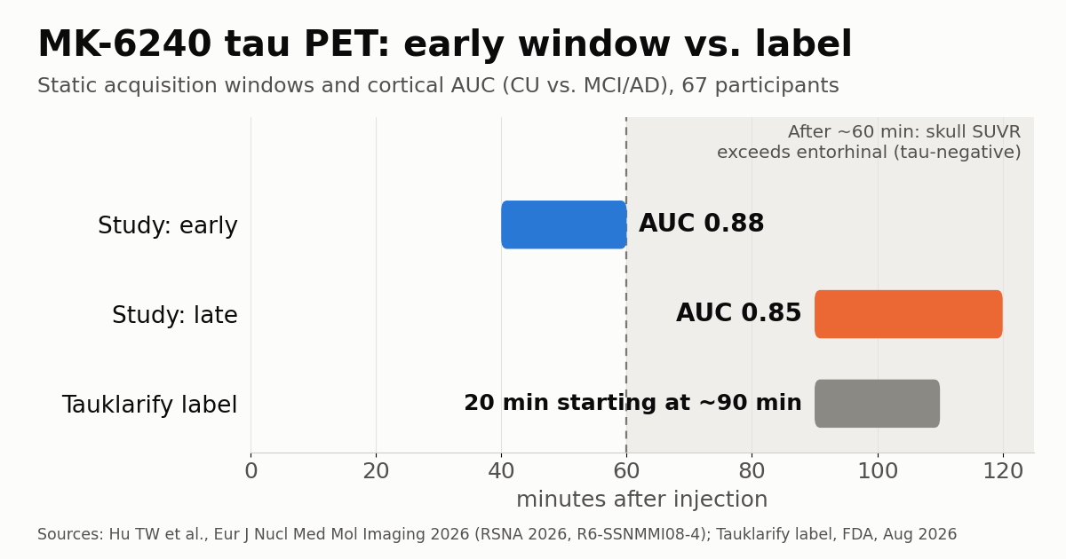
An early F-18 MK-6240 tau PET acquisition, 40 to 60 minutes after injection, separated cognitively unimpaired people from patients with mild cognitive impairment (MCI) or Alzheimer’s disease about as accurately as the late 90- to 120-minute window, with less contamination from the skull and meninges. The work, from Weill Cornell Medicine in New York, will be presented at RSNA 2026, three months after the FDA approved the tracer with an imaging protocol that starts at roughly 90 minutes.
Tsung-Wei Hu, PhD, of Weill Cornell’s Brain Health Imaging Institute, presents the results on December 3 (session R6-SSNMMI08-4). The same dataset was published in June in the European Journal of Nuclear Medicine and Molecular Imaging.
What the Weill Cornell team measured
The analysis is retrospective and within-subject. Each of the 67 participants (cognitively unimpaired, MCI and Alzheimer’s) had MK-6240 PET frames in both an early (40 to 60 minutes) and a late (90 to 120 minutes) window. The authors computed standardized uptake value ratios (SUVR) in the cerebral cortex and the Braak staging regions, with cerebellar cortex as the reference, and graded and quantified skull and meningeal uptake. The headline findings:
- Early and late SUVRs correlated at r ≥ 0.97, with near-identical cohort distributions.
- Cortical SUVR separated cognitively unimpaired participants from those with MCI or Alzheimer’s with an area under the ROC curve (AUC) of 0.88 in the early window and 0.85 in the late one.
- In tau-negative participants, skull SUVR exceeded entorhinal SUVR from roughly 60 minutes onward.
- In simulated PET-to-MRI misregistration, late entorhinal SUVR varied about four times more than early SUVR (standard deviation 0.071 vs. 0.017).
“The 40-60-minute window provides discrimination comparable to 90-120 minutes while reducing extracerebral contamination and improving robustness,” the group wrote, arguing for shorter static protocols in both clinical and research use.
Correction: MK-6240 is no longer under FDA review
The RSNA abstract, as reproduced by AuntMinnie, calls MK-6240 a tracer “currently under FDA review.” That is out of date. The FDA approved florquinitau F 18, the generic name for MK-6240, in August 2026 under the brand Tauklarify, and Lantheus announced the decision on August 14, as we reported in FDA approves Lantheus tau PET agent for Alzheimer’s. The wording comes from the group’s paper, published online on June 13, two months before the approval.
The label sets the protocol: 185 MBq (5 mCi) as an intravenous bolus and a 20-minute PET image starting approximately 90 minutes after injection. Lantheus said that, for now, it will keep supporting pharmaceutical partners’ Alzheimer’s therapy programs while it assesses the path to broader commercial availability.
Why the label settled on 90 minutes
In the FDA’s multidisciplinary review, reviewers noted that brain signal stabilizes by about 40 to 60 minutes, that SUVR referenced to cerebellar cortex plateaus after roughly 60 minutes, and that images acquired after 90 minutes are expected to perform like those acquired between 60 and 90 minutes. The 90- to 110-minute window was accepted because it was used in the two pivotal studies (CE-20-01 and CE-23-01) that established efficacy. In other words, the label reflects how the trials were run, not evidence that waiting longer yields a better scan.
The label also anticipates the problem the Weill Cornell group quantified: it tells readers to treat signal at the junction of the skull base and the meninges as off-target, whether isolated hotspots or a ring-like pattern, and to leave it out of the read. For comparison, flortaucipir (Tauvid), the first tau PET agent approved in the U.S. in 2020, is labeled at 370 MBq with a 20-minute image starting about 80 minutes after injection.
Braak staging, SUVR and signal outside the brain
Tau tangles in Alzheimer’s disease follow the route described by Braak staging: they start in the transentorhinal and entorhinal cortex (stages I and II), spread to limbic structures (III and IV) and finally reach the neocortex (V and VI). The entorhinal cortex, which flags early disease, sits close to the skull base. Because SUVR divides target-region uptake by uptake in a reference assumed free of tangles, signal that spills in from bone or meninges through partial-volume effects inflates the numerator and can push a negative scan toward positivity.
Meningeal uptake of MK-6240 was already documented in the literature. On the other hand, the tracer shows minimal choroid plexus binding, a known confounder of medial temporal reads with flortaucipir. In the HEAD study, published in The Lancet in 2026 with 682 participants who had both scans, MK-6240 reached an AUC of 0.93 versus 0.86 for flortaucipir in distinguishing Alzheimer’s from other causes of cognitive impairment. Non-Alzheimer tauopathies remain outside the indication and have their own tracers in testing, such as the 18F-OXD-2314 agent studied in chronic traumatic encephalopathy.
What a shorter window changes for nuclear medicine
Table time does not change: the image takes 20 minutes either way. What shrinks is the wait, since the exam ends about an hour after injection instead of nearly two. For patients with cognitive impairment that difference matters, and it frees up uptake rooms and PET/CT schedules. Shorter protocols are a broad trend in PET, as shown by work on total-body PET delivering usable images in one minute.
The label carries another scheduling detail: CYP1A2 inducers, including tobacco smoking, should be avoided for at least seven days before injection. In Brazil, Anvisa has registered the anti-amyloid drugs donanemab (April 2025) and lecanemab (December 2025), but the U.S. approval does not carry over to Latin America: clinical use of the tracer there depends on local registration.
Limitations and what to watch
The study is retrospective and small, and the abstract does not give the split between diagnostic groups. The AUC difference (0.88 vs. 0.85) supports comparable performance, not superiority. For the diagnostic analysis, the reference standard was clinical diagnosis, without neuropathological confirmation. And the analysis is quantitative, with MRI-defined regions, whereas the label relies on a binary visual read by readers trained on 90- to 110-minute images; the early window has not yet been validated with that read method.
There is also a kinetic caveat: earlier dynamic studies favored late windows because, in high-binding regions, late SUVR agrees better with kinetic models. In clinical trials and longitudinal follow-up, mixing windows between scans would introduce bias. Researchers at the same institute showed in 2024 that MK-6240 kinetic modeling is feasible with dynamic acquisitions as short as 40 minutes in cognitively normal volunteers. The next things to watch are multicenter validation with visual reads and whether the manufacturer seeks to add a shorter window to the label.
Source: AuntMinnie — Early F-18 MK-6240 tau PET scans feasible. Original paper: Hu TW et al., Eur J Nucl Med Mol Imaging, 2026; Tauklarify prescribing information (FDA, 2026).