

Measuring how much the brain moves with every heartbeat predicts the outcome of decompression surgery in Chiari malformation type I better than the anatomical measure used for decades — cerebellar tonsillar descent. That is the conclusion of an Emory University study published in the Journal of Neurosurgery, which paired phase-contrast MRI to quantify cerebrospinal fluid flow with a displacement technique called cine DENSE to measure tissue motion on the order of micrometers.
The clinical problem: operating on the people who will actually improve
Chiari malformation type I is defined by herniation of the lower cerebellum — the cerebellar tonsils — at least 5 mm through the foramen magnum into the spinal canal. It is a congenital condition, but many patients only develop symptoms in adulthood: occipital headache worsened by exertion, dizziness, impaired balance, dysphagia.
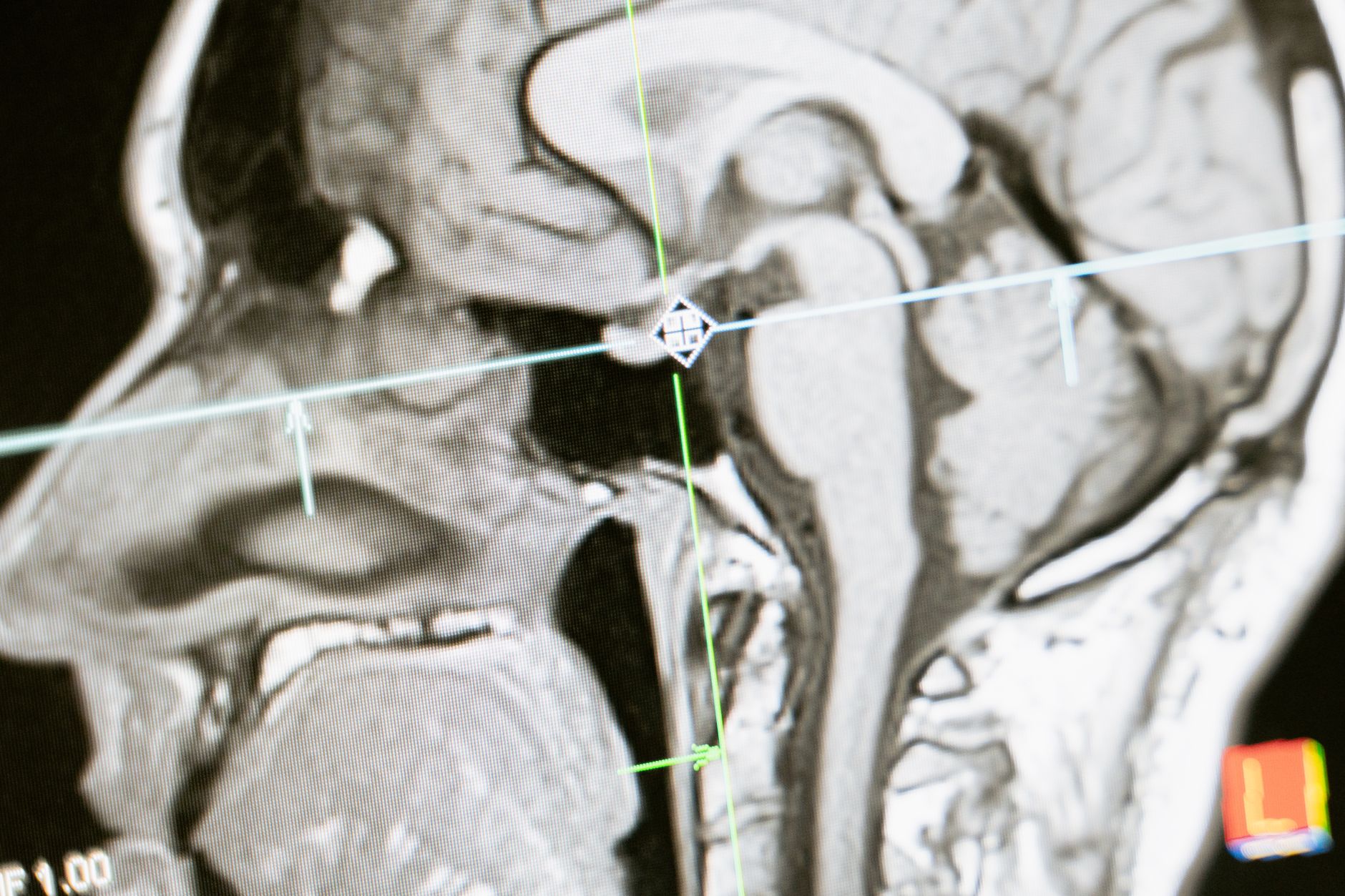
Posterior fossa decompression (PFD) improves symptoms in roughly 75% of cases. The other side of that statistic is what bothers clinicians: one in four patients undergoes a procedure carrying real risks — CSF leak, meningitis, pseudomeningocele — without the expected benefit. And the imaging criterion practice leans on to decide, tonsillar descent in millimeters, has a track record of inconsistent results as a predictor.
What the study measured
The team enrolled 108 participants aged 19 to 70, of whom 96 entered the analysis after exclusions. Sixty-one underwent posterior fossa decompression and 48 completed postsurgical imaging. A group of 25 healthy volunteers supplied reference values for brainstem and cerebellar motion. All scans ran on a Siemens Prisma Fit 3.0-T system with a 20-channel head/neck coil.
Two functional measures were extracted. The first is CSF stroke volume, obtained by phase-contrast MRI in an axial plane perpendicular to the spinal cord at the C2 or C6 level — 1.2 × 1.2 × 5 mm³ resolution, TE/TR of 6/21 ms, VENC of 15 cm/s, 25 frames per cardiac cycle, about 1 minute 48 seconds of acquisition. The volume is half the absolute cranial and caudal flow integrated over the cycle:
$$V_{\text{CSF}} = \tfrac{1}{2} \int_{0}^{T} \left| Q(t) \right| \, dt$$
The second is brain tissue motion, measured with cine DENSE (displacement encoding with stimulated echoes) in a midsagittal plane, at 30 to 40 ms temporal resolution, 0.9 × 0.9 mm pixels, 8 mm slice thickness and encoding in both anterior-posterior and cranial-caudal directions. What makes the technique work is the relationship between phase and displacement: for an encoding frequency $k_e$, displacement $u$ is recovered from the measured phase $\phi$ as
$$u = \frac{\phi}{2\pi k_e}$$
With the protocol’s $k_e$ of 0.6 cycles/mm, a full phase cycle corresponds to roughly 1.67 mm of displacement. That is what makes submillimeter motion measurable with confidence: 187 µm of brainstem displacement is about one tenth of a phase cycle, something no morphological measurement would capture.
The numbers before and after surgery
CSF flow increased after decompression: stroke volume went from 0.49 ± 0.24 mL per cycle to 0.63 ± 0.28 mL per cycle, a 28.9% rise (p = 0.014). In the paired analysis restricted to surgical patients, the average increase was 22.6% (p = 0.057). Peak velocity did not change meaningfully (5.81 versus 5.51 cm/s; p = 0.178) — meaning surgery widens the volume that passes, not the speed at which it passes.
Tissue motion fell, moving toward healthy control values. In the brainstem, maximum displacement was 187 ± 64 µm before and 162 ± 53 µm after, a paired reduction of 17.3% (p = 0.002); the healthy volunteer reference is 117 µm. In the cerebellum the change was larger: from 142 ± 69 µm to 91 ± 36 µm, a 45.2% reduction (p < 0.001), against 67 µm in controls. The cranial-caudal component accounted for most of the drop.
But the study’s central finding lives in the correlations. Presurgical tonsillar descent did not relate to postsurgical improvement in CSF flow (R = 0.059; p = 0.767) — essentially noise. The presurgical functional measures predicted well: prior CSF flow against flow improvement gave R = −0.518 (p = 0.005), prior brainstem motion against its reduction gave R = −0.638 (p < 0.001), and prior cerebellar motion against its reduction reached R = −0.878 (p < 0.001). The negative sign is exactly what you would expect: those most deranged beforehand gained the most.
“The measurement of neural dynamics, such as brain motion and CSF flow, rather than static markers, such as tonsillar descent, are a new approach to understanding the pathophysiology of brain disease and represent a new method to improve patient treatment options,” summarized John Oshinski, professor of radiology and imaging sciences and of biomedical engineering at Emory, who led the work with neurosurgeon Daniel Barrow. First author Grace McIlvain is now an assistant professor of biomedical engineering and radiology at Columbia University.
Why this matters to radiology, not just neurosurgery
The transferable concept is swapping a static marker for a dynamic one. Chiari is a teaching case because anatomy measures the presumed cause — cerebellum obstructing the foramen magnum — while physiology measures the consequence that produces symptoms, namely altered pressure and tissue motion. When the two diverge, outcome follows physiology.
The pattern shows up across neuroimaging. We have covered how advanced MRI detects functional brain changes in long COVID that conventional structural imaging does not show, and how AI models extract multiple diagnostic signals from a single brain MRI — including parameters no radiologist measures by eye. Same direction: pull quantitative, non-morphological information out of the MRI.
There is an operational message too. The two sequences add up to under five minutes of acquisition — 1 min 48 s of phase contrast and about 3 minutes of DENSE — on an ordinary 3 T scanner. This is not research demanding exotic hardware. The bottleneck lies elsewhere: spiral DENSE with peripheral pulse gating is not a product sequence on most installed systems, and phase-to-displacement post-processing needs dedicated tooling. Services already running cardiac MRI with tissue tagging are closer to reproducing this than they might assume.
Limitations and the next step
The authors are explicit: the data “do not yet establish a clinical standard for deciding surgery.” The series comes from a single surgical center and a single surgeon with 39 years of post-fellowship experience, which limits generalization. Not every participant had complete imaging at both timepoints — there were 70 usable DENSE and 67 phase-contrast scans before surgery, but only 31 and 28 afterward, with just 16 patients having both measures postoperatively.
There are honest mechanistic limits as well. What drives the correlation between brain motion and CSF flow is unknown, and flow is affected by factors beyond the cardiac cycle — respiration and neural activity among them. The link to long-term clinical outcome is not established: the correlation between presurgical cerebellar motion and the Chiari Clinical Outcome Scale sat at the edge of significance (R = 0.308; p = 0.053). Translated: functional imaging predicts physiological change well, and still has to prove it predicts patient improvement.
The next step is already underway — a larger, blinded clinical trial recruiting additional sites. Until then, the practical message is more modest and more useful than the headline: in Chiari patients with a borderline surgical indication, measuring CSF flow and tissue motion adds information tonsillar descent does not provide. Worth remembering that neuroimaging decisions have been shifting fast — we saw it when rapid MRI began being argued for in place of CT in pediatric head trauma — and that the MRI suite keeps moving closer to the operating room, as with open intraoperative MRI for glioma.



