

An AI model trained on adult radiographs can be adapted to newborns without any hospital sharing patient data. That is the core finding of an international study published on October 13, 2026, in Nature Biomedical Engineering: using continual learning across 30 neonatal intensive care units (NICUs) in 20 countries and territories, the error in locating the endotracheal tube tip and carina fell to 0.67 cm, versus 1.29 cm with single-hospital fine-tuning and 4.30 cm when the adult model was applied unchanged.
The problem: adult AI does not read children well
The authors point out that only about 17% of commercial AI models have FDA clearance for pediatric use, and that children make up less than 1% of public medical imaging datasets. Most published pediatric models come from a single center, few are externally validated, and many lose accuracy when they move to another institution.
As general background, the reasons for the performance drop are familiar to pediatric radiologists. A preterm chest has very different proportions from an adult one, structures are measured in millimeters, films are taken at the bedside inside an incubator with leads, electrodes and catheters overlapping the anatomy, and exposure protocols are tuned for low dose. Disease prevalence shifts as well. An algorithm that has never seen that combination can miss by several centimeters. The same issue frames other pediatric imaging work we have covered, such as the case for rapid MRI in pediatric head trauma and the push for safer pediatric MRI protocols.
How the study was done
The work was led by Vishwanatha M. Rao and supervised by Pranav Rajpurkar of the Department of Biomedical Informatics at Harvard Medical School, with dozens of coauthors from sites including Stanford, Great Ormond Street Hospital in London, Osaka Metropolitan University, the University of Cape Town, and the AI committee of the Colombian Association of Radiology, along with a university hospital in Manizales, Colombia.
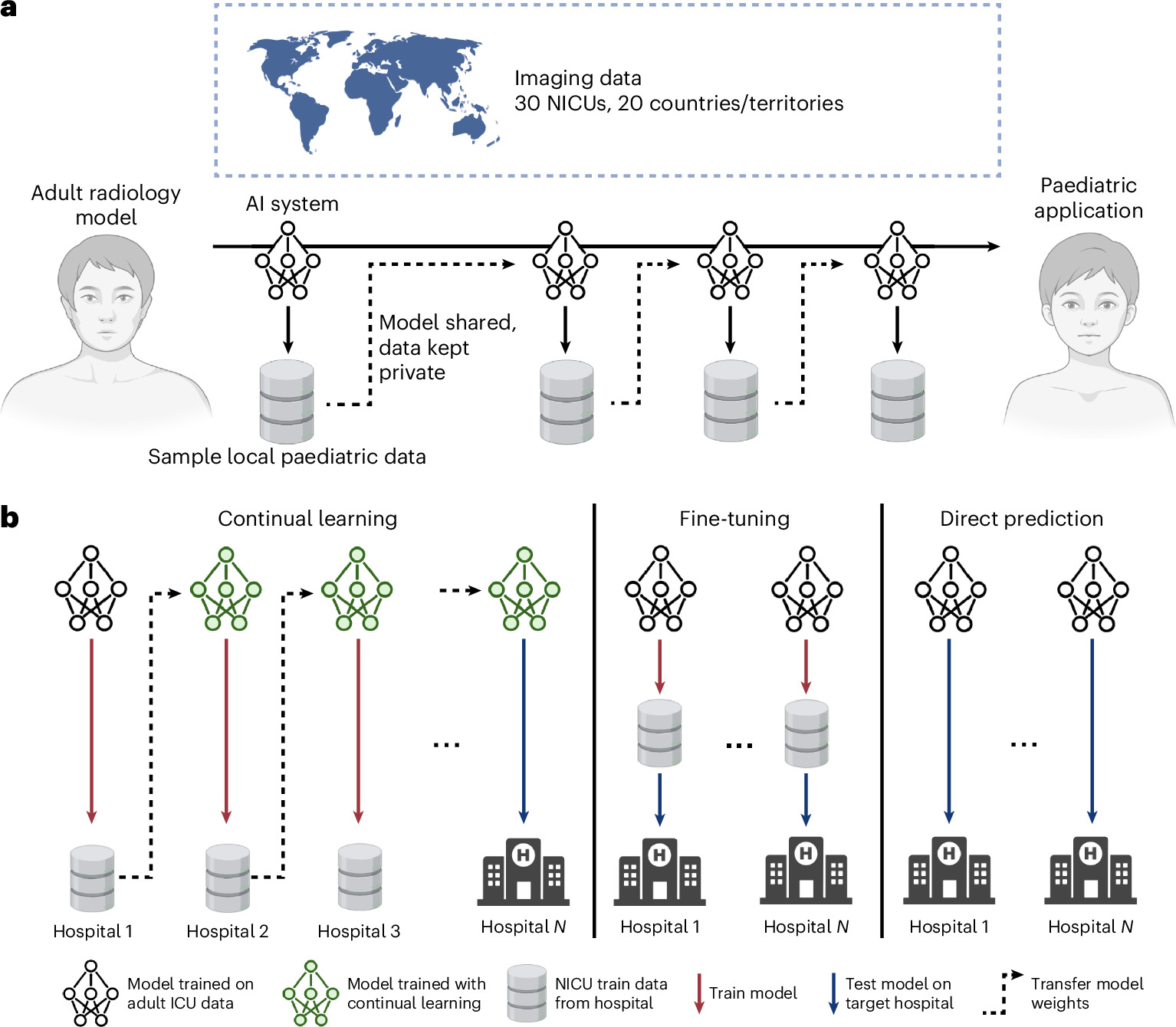
The chosen task was endotracheal tube (ETT) position assessment, for which FDA-cleared tools already exist in adults but no commercial solution exists for neonates. The starting point was CarinaNet, a RetinaNet-based object detector with a ResNet-50 backbone, previously trained on 1,890 adult ICU radiographs to locate the tube tip and carina.
Each NICU contributed about 100 radiographs, roughly half with well-placed and half with malpositioned tubes. Fifty images were used for local training and the rest (43 to 53 per site) for testing. The final cohort included 2,965 radiographs from 2,960 patients, with a median age of 2 days, mean birth weight of 2,195 g and mean gestational age of 32.9 weeks, a predominantly preterm population.
Under continual learning, the first hospital receives the adult model, retrains it on its 50 images and passes only the weights to the next site, and so on. The authors compared this with two realistic alternatives: applying the adult model as is (direct prediction) or retraining it only on the target hospital’s data (fine-tuning). For each of the 30 sites, the evaluated hospital was left out of the training sequence, and results were averaged over 10 random hospital orderings. Experiments were run centrally to simulate decentralized execution.
Key results
- Tube tip: median error of 2.35 cm with direct prediction, 0.94 cm with fine-tuning and 0.40 cm with continual learning.
- Carina: 1.62 cm, 0.27 cm and 0.20 cm, respectively.
- Combined error: 0.67 cm with continual learning; improvement at 29 of 30 hospitals, statistically significant at 27.
- Extreme errors (>5 cm): 2.6% of cases with continual learning, 8.2% with fine-tuning and 37.4% with direct prediction. The threshold was chosen because 5 cm roughly equals the full tracheal length of many neonates.
- Triage of deep intubations: precision and recall among the 15 most concerning films improved over fine-tuning at 27 of 30 sites (significant at 18), with precision above 80% at the best-performing hospitals and fewer false positives at 18 sites.
Three further analyses explain the result. The model did not forget hospitals seen early in the sequence; their performance actually improved by a mean 0.48 cm by the end. Adult pretraining mattered: starting from a generic detector with no radiograph exposure did worse at 25 of 30 sites. And gains grew with the number of participating hospitals, with no plateau. On the other hand, adding a 31st site with low-resolution images worsened combined error (0.75 cm versus 0.67 cm), showing that each participant’s data quality counts.
Why the approach matters for hospitals and vendors
Technically, the method is single-pass serial weight transfer, the so-called “travelling model”. Its advantage over federated learning is simplicity: no central server, no synchronized communication rounds and no need for matching infrastructure. The authors envision a training container shipped to each hospital, run by local IT against preset acceptance criteria, with a versioned ledger that allows rollback if a regression appears. Clinicians would only need to curate and annotate a few dozen studies.
For health systems in Latin America and other regions with strict privacy laws and small NICU volumes, the model is attractive because patient images never leave the building. The participation of Colombian centers shows that hospitals outside the usual North American and European networks can join such consortia. It is also a reminder that pediatric AI remains scarce on regulatory lists: the growth in clearances we analyzed in FDA radiology AI grows 42% remains adult-centric, and real deployments face their own bottlenecks, as shown in real-world radiology AI.
Limitations and next steps
The study is retrospective and the authors explicitly call it a proof of concept. Datasets were artificially balanced at about 50% malpositioned tubes, which does not reflect real prevalence; pixel-to-centimeter conversion relied on estimated tube width because DICOM pixel spacing was inconsistent across sites; and tube-to-carina distance is a weak proxy for high tubes, which are defined relative to the first thoracic vertebra. Decentralization was simulated, and a model that changes at every hospital complicates regulatory oversight, since each update must be reconciled with FDA processes and local review boards. In extremely preterm infants the acceptable margin for tube position is even narrower, so dedicated prospective validation is needed.
The proposed next step is to integrate the model into clinical workflows at a subset of participating NICUs to measure time to radiographic confirmation of malpositioned tubes, clinician agreement with model flags and the effect on reintubation rates. If it holds up, the same recipe could be applied to other pediatric tasks where adult AI exists but nothing has been built for children.
Source: Rao et al., Nature Biomedical Engineering 2026 (doi:10.1038/s41551-026-01812-6), as reported by Health Imaging



