
Social isolation and loneliness, two recognized risk factors for dementia, are linked to different but partly overlapping patterns of brain change on MRI and PET in middle-aged and older adults. That is the conclusion of a systematic review of 42 studies by researchers at Deakin University in Geelong, Australia, published October 28, 2026, in Alzheimer’s & Dementia.
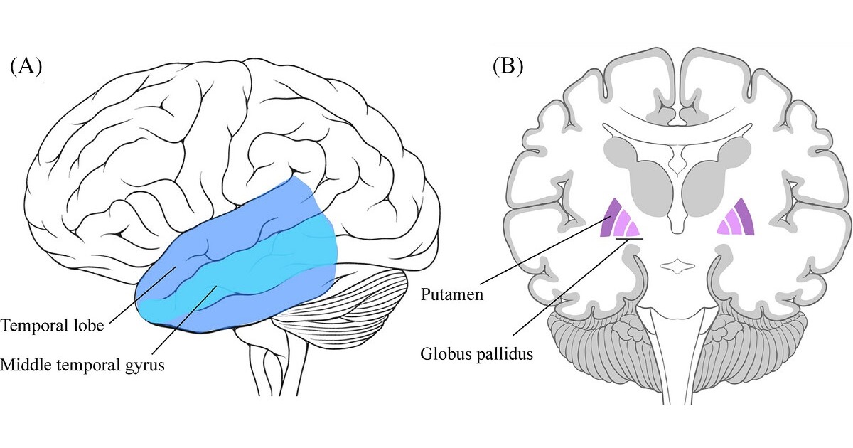
Across the literature, social isolation — the objective state of having few contacts and little social participation — was more often associated with temporal regions such as the middle temporal gyrus. Loneliness, the subjective sense of having fewer meaningful relationships than one wants, tracked more closely with subcortical structures, notably the putamen and globus pallidus. Both shared findings in limbic regions, greater white matter hyperintensity volume and elevated C-reactive protein.
How the Deakin team built the review
The work, led by PhD candidate Jade Guarnera with Helen Macpherson as senior author, starts from a measurement problem. Researchers often use the two terms interchangeably, or apply a questionnaire designed for one construct when studying the other. That blurs two phenomena that may run through different biological pathways: a person can live alone without feeling lonely, while someone surrounded by family can report intense loneliness.
The team searched PubMed, PsycInfo, Medline ProQuest and Embase for studies published from 2010 to 2025 that related social isolation or loneliness to neurobiological outcomes in samples with a mean age of 50 or older. Eligible outcomes covered structural and functional MRI, diffusion tensor imaging (DTI), amyloid and tau PET, cerebrospinal fluid, and blood-based inflammatory markers such as C-reactive protein (CRP). The final set held 42 studies: seven on social isolation, 26 on loneliness and nine that measured both.
“We currently lack a strong understanding of the mechanisms that might explain this risk, particularly the underlying neurobiology,” the authors wrote. To their knowledge, it is the first systematic review of the neurobiological correlates of both constructs in middle-aged and older adults.
Temporal cortex, basal ganglia and shared limbic ground
The paper’s main figure shows only associations that were exclusive to one construct and replicated in at least two studies. For social isolation, that means the temporal lobe and, within it, the middle temporal gyrus, a region involved in semantic processing and social cognition. For loneliness, it means the putamen and globus pallidus, basal ganglia nuclei that take part in circuits for movement, habit formation and reward processing.
The overlap includes the hippocampus, amygdala, thalamus and fusiform gyrus, which the authors group as limbic regions. The hippocampus is among the earliest structures to atrophy in Alzheimer’s disease, and the amygdala is central to emotional processing. The authors point out that several of the implicated regions are vulnerable in Alzheimer’s disease and related dementias, suggesting possible pathways from poor social health to neurodegenerative risk.
Beyond gray matter, both constructs were associated with greater white matter hyperintensity or lesion volume and with higher CRP, a marker of systemic inflammation. The pattern fits other recent work tying social conditions to brain imaging, including a study that linked neighborhood deprivation to faster brain aging on MRI.
What each imaging measure actually captures
For imaging readers, it helps to be precise about what each technique reports. T1-weighted structural MRI allows the brain to be segmented so that gray matter volume and cortical thickness can be measured; the middle temporal gyrus, putamen and hippocampus findings come from this kind of analysis. Functional MRI follows the BOLD signal, which reflects changes in blood oxygenation, and is mostly used to estimate connectivity between regions, as in a recent fMRI study of music and recovery from acute stress.
DTI measures how water diffuses along fiber bundles and yields indices such as fractional anisotropy, used as proxies for white matter microstructural integrity. White matter hyperintensities show up as bright areas on T2 and FLAIR images and usually reflect small vessel disease, a common finding in older adults that is associated with stroke and cognitive decline. Amyloid and tau PET use radiotracers that bind to the plaques and tangles that define Alzheimer’s pathology; the recent FDA approval of a new tau PET agent broadens clinical access to the second of those tools.
Association, not causation: limits of the evidence
The authors acknowledge that substantial methodological heterogeneity and inconsistent measurement of the two constructs limit what can be concluded. A systematic review of this kind pools observational studies with different samples, acquisition protocols and analysis pipelines. Judging from the abstract and the figure, the synthesis rests on how consistently a region appears across studies rather than on a pooled effect size.
Direction is another open question. When a scan and a questionnaire are collected at the same time, there is no way to tell which came first: loneliness may contribute to brain change, but early neurodegeneration can also lead people to withdraw socially. Depression, hearing loss, socioeconomic status and physical activity all intersect with both constructs and can confound the association.
The evidence base is also uneven. Counting exclusive and combined studies, 35 assessed loneliness but only 16 assessed social isolation, so the isolation map rests on fewer data. None of these findings allows a clinician to infer a patient’s social situation from a scan, or to estimate an individual’s dementia risk.
What changes for neuroimaging research and practice
For routine reporting, nothing changes: the differences described are group averages that cannot be seen when reading a single study. The value of the review lies in shaping the next generation of research. The authors call for validated multi-item loneliness scales, more fMRI and broader white matter metrics in older samples, and designs that analyze isolation and loneliness within the same model so that results can be compared. “Moreover, the development of a multidimensional, social isolation-specific measure should be prioritized as the social health field advances,” they concluded.
The work adds to a broader effort to pull early signs of cognitive decline out of routine MRI, such as tight sulci linked to cognitive decline and an MRI signature that predicts progression from MCI to early-onset dementia. Cohorts that already acquire brain MRI, including those in fast-aging populations in Latin America, could add validated social measures that keep isolation and loneliness apart and test whether these patterns hold in different socioeconomic settings. The 2024 update of the Lancet Commission on dementia already lists social isolation among the modifiable risk factors.
Source: AuntMinnie — MRI, PET reveal distinct brain changes in loneliness and social isolation. Original paper: Guarnera J et al. The underlying neurobiology of social isolation and loneliness in middle-older adults: A systematic review. Alzheimer’s & Dementia, 2026.