

PSMA PET/CT-guided biopsy found nearly twice as much clinically significant prostate cancer as systematic biopsy in men whose MRI was negative or equivocal. In the prospective FUPERMAN study from Bologna, Italy, published in the Journal of Nuclear Medicine (JNM), PET-targeted biopsy detected significant disease in 37% of 63 patients versus 21% for systematic biopsy; combining both techniques raised the yield to 41%. The Society of Nuclear Medicine and Molecular Imaging (SNMMI) highlighted the work in an October 5 news release.
How FUPERMAN was designed
The study was led by Andrea Farolfi of IRCCS Azienda Ospedaliero-Universitaria di Bologna, with Lorenzo Bianchi and Stefano Fanti among the coauthors, and appeared in the September issue of JNM (vol. 67, no. 9, pp. 1392-1400) as an open-access paper. The acronym stands for fusion PSMA PET in MRI-negative men. Between March 2023 and July 2025 the team enrolled 63 consecutive men with PI-RADS 2 (51 patients) or PI-RADS 3 (12) multiparametric MRI who still had a clinical reason for suspicion: abnormal digital rectal exam, PSA density of at least 0.1 ng/mL/mL, PSA of 4 ng/mL or higher, family history, or a germline BRCA1/2 mutation. PSA was rechecked four to six months after MRI to confirm it remained abnormal. About a third of the men had a prior biopsy.
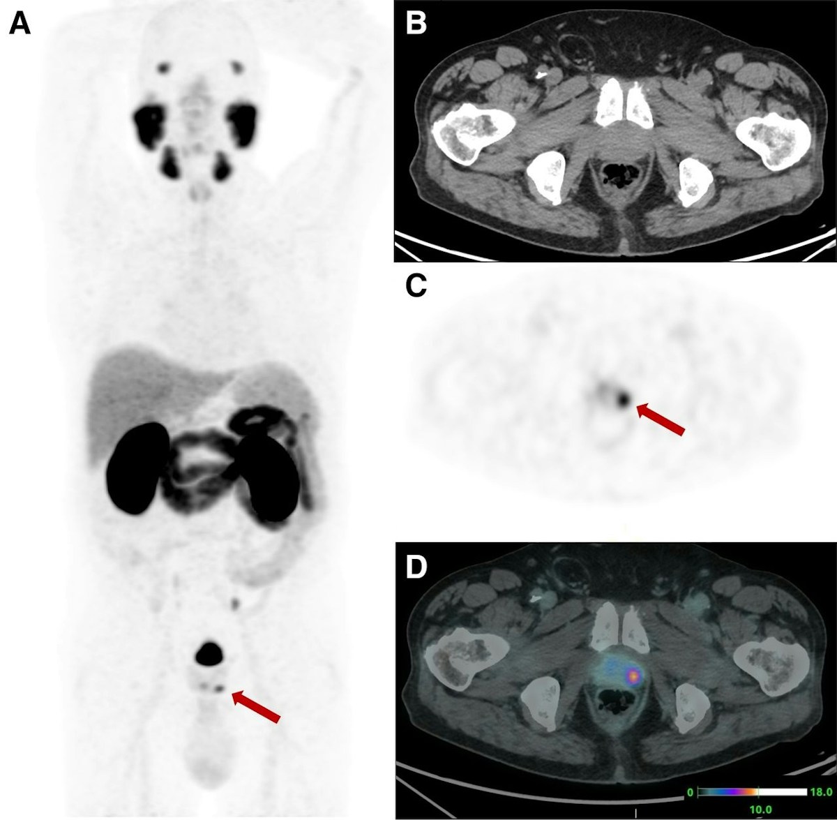
All underwent [68Ga]Ga-PSMA-11 PET/CT (1.8 to 2.2 MBq/kg, imaging at 60 minutes plus a delayed pelvic scan at 90), read by two nuclear medicine physicians using the PRIMARY score; scores of 3 to 5 counted as positive. Then came an outpatient transperineal biopsy under local anesthesia by a single urologist: at least eight systematic cores for everyone and, in PET-positive men, at least three cores per suspicious area, aimed with real-time fusion. Prostate and lesion contours drawn on CT and PET were automatically overlaid on the 3D transrectal ultrasound volume (UroFusion software on an Esaote MyLab X9 platform). PI-RADS 3 cases also received MRI-targeted cores.
The results
Overall, 26 of 63 men (41%) had clinically significant cancer, defined as ISUP grade group 2 or higher. The rate was essentially the same for PI-RADS 2 (41%) and PI-RADS 3 (42%), which says a lot about the residual risk in this selected group. PET was positive in 44 patients (70%) and flagged 25 of the 26 significant cancers.
As a diagnostic test, PSMA-targeted biopsy reached 96% sensitivity, 49% specificity, a 57% positive predictive value and a 95% negative predictive value. Its detection advantage over systematic biopsy (37% vs. 21%) was statistically significant (p=0.021). Agreement between the two approaches was poor (kappa 0.116): each found cancers the other missed, and PET-targeted cores showed a higher ISUP grade than systematic cores in 17 patients (27%). Three significant cancers were missed by targeted biopsy, two of them invisible on PET. The scan also revealed pelvic nodes or metastatic disease in four men (6%).
Uptake intensity carried its own information. Median SUVmax was 4.3 in men without significant disease and 14.8 in those with ISUP 2 to 5, and the ROC area under the curve for SUVmax (0.847) beat that of PSA density (0.698). A threshold of 11.4 gave 100% specificity at 58% sensitivity, very close to the cutoff of 12 reported in the PRIMARY study. On multivariable analysis, age, prostate volume and SUVmax were independent predictors.
Dynamic total-body PET: an exploratory signal
Eleven patients also had a 60-minute dynamic acquisition on the uEXPLORER, a total-body PET/CT with a 194 cm axial field of view. Using an image-derived input function from the descending aorta, segmented automatically with TotalSegmentator, the authors ran Patlak analysis and two-tissue compartment models. Every area with irreversible uptake kinetics proved to be significant cancer at histology, and the net influx constant Ki separated true lesions from false positives better than SUVmax (AUC 0.97 vs. 0.92). In PRIMARY 3 lesions, the most ambiguous group, the k3 rate constant discriminated where SUV could not. With only 11 patients, the authors frame this as proof of concept, not a clinical tool.
Where FUPERMAN fits in the pre-biopsy PSMA debate
This study is distinct from, but complementary to, the news that the FDA granted Fast Track to pre-biopsy PSMA PET plus MRI. The randomized PRIMARY2 trial and Telix’s BiPASS program ask whether PET can safely let men avoid biopsy. FUPERMAN asks a different question: when a biopsy is going to happen anyway, does PET help aim it? All 63 men had systematic biopsy, and PET added targets on top. The technique differs too. In the original PRIMARY study, urologists targeted using static key images, a cognitive approach; here, PET regions feed a real-time ultrasound fusion system, which should reduce operator dependence.
The authors argue that PET should complement rather than replace systematic sampling, precisely because of the poor agreement between techniques and the modest specificity. That is a more conservative stance than PRIMARY2’s and consistent with meta-analyses warning that a high negative predictive value alone is not enough to skip biopsy.
Practical implications for imaging services
For radiologists, the message is that a PI-RADS 2 or 3 report in a man with persistent PSA elevation and high PSA density does not close the case. These patients often end up with repeated random biopsies of low yield. In a well-designed pathway MRI remains the entry point, and even that is not a given: in the United States, pre-biopsy MRI is still used in only a third of cases. PSMA PET would come in as a problem-solving step, with a structured report combining PI-RADS, PRIMARY score and SUVmax.
In Brazil and much of Latin America, gallium-68 PSMA PET is offered by private and academic nuclear medicine services, mainly for staging and biochemical recurrence. Pre-biopsy diagnostic use sits outside the more established indications and is generally not covered. PET-ultrasound fusion also requires software that can import PET data and a joint workflow between nuclear medicine, radiology and urology, which is more realistic in referral centers. For radiation oncology, a dominant lesion located on two modalities is useful for planning and for weighing a focal boost, close to the debate on the role of MRI in prostate cancer staging.
Limitations and next steps
This is a small single-center study in a deliberately enriched high-risk population, which explains the 41% prevalence of significant disease, well above what is expected in unselected PI-RADS 2-3 men. The authors caution that the findings do not apply to population screening. Systematic biopsy used a limited number of cores rather than saturation mapping, which may have tilted the comparison toward PET. The interval between MRI and biopsy was relatively long. And 49% specificity means many targets without significant cancer. The work was funded by the Italian Ministry of Health; Fanti and Farolfi disclosed honoraria and travel support from United Imaging, maker of the uEXPLORER.
Even so, FUPERMAN adds a piece to the picture that has been building since PRIMARY and that drove part of the conversation at the 2026 SNMMI annual meeting: PSMA PET is moving beyond staging and into diagnosis. Confirmation will have to come from larger multicenter studies, including longer follow-up of PRIMARY2 itself.
Source: AuntMinnie and Farolfi et al., Journal of Nuclear Medicine 2026



