A New Generation of Radiotherapy Simulation
Philips unveiled on May 26, 2026 the 4D MR-RT, an MRI solution designed specifically for radiotherapy simulation of abdominal tumors affected by respiratory motion. The platform combines respiratory phase-resolved 4D acquisition, multiple image contrasts in a single sequence and free breathing — all in a workflow built for the radiation oncologist, not just the diagnostic radiologist.
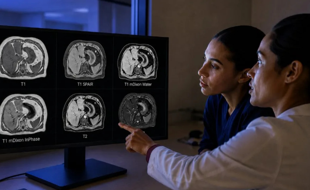
The technical problem the product addresses is long-standing. In liver, pancreas and adrenal treatments, breathing can shift the target by up to 3 cm in the cranio-caudal axis, and traditional methods — breath-hold, abdominal compression, respiratory gating — either undermine reproducibility or compromise patient comfort. The 4D MR-RT proposition is to capture real motion during free breathing and use that information to inform both target margins and treatment phase selection.
How the Acquisition Works
The system relies on Philips’s SmartSpeed technology to distinguish inhale and exhale phases during free breathing. Reconstruction produces up to 10 respiratory phases, generating time-resolved volumes. Phases can be used individually or combined to generate mid-ventilation and mid-position images, which serve as reference data for target and organ-at-risk contouring in linear accelerator-based planning.
The differentiator over conventional 4D-CT is soft-tissue contrast. The solution incorporates T1- and T2-weighted imaging, with optional fat suppression, in a single sequence. That allows visualization of liver, pancreas and adjacent structures without switching machines or repeating acquisitions at a different point in the respiratory cycle.
Why It Matters for Practice
Abdominal radiotherapy simulation has always carried a trade-off: CT provides direct electron density for dose calculation but limited soft-tissue contrast; MR offers excellent contrast but requires synchronized workflows and multiple acquisition stations. The 4D MR-RT consolidates those steps into a single session, reducing simulation time and inter-operator variability.
For services already running IGRT in abdominal SBRT — as we covered in our ASTRO 2026 presidential symposium coverage — that quality gain translates into tighter margins, lower dose to organs at risk and potential expansion of SBRT indications to liver and pancreas. The system also integrates with MR-Linac workflows, although Philips positions the product primarily for simulation on conventional LINACs.
Regulatory Status and Availability
Philips states that the 4D MR-RT has received CE Mark certification for clinical use in Europe and FDA 510(k) clearance for clinical use in the United States. For other markets, the next step is local regulatory registration, which typically takes 12 to 18 months for dedicated MRI solutions, depending on the clinical documentation submitted. Academic centers already operating Philips platforms may become early adopters.
Ioannis Panagiotelis, business leader for magnetic resonance at Philips, positioned the product in an official statement: “With 4D MR-RT, Philips is advancing MR simulation, bringing greater confidence, consistency and accuracy to treatment planning, while supporting a more comfortable and reproducible patient experience.”
Implications for the RT-MRI Market
The launch puts Philips in direct competition with Siemens Healthineers and GE HealthCare, both also investing in dedicated MRI for radiotherapy. Siemens has the MAGNETOM Vida ecosystem with RT configuration, and GE HealthCare recently launched the MIM ComboTherapy GYN with a heavy 4D imaging component. In our analysis of the MIM ComboTherapy GYN, we explored how these workflows are being designed to reduce inter-observer variability.
For international markets, the implication is direct: radiotherapy services that rely on conventional CT simulation for abdominal tumors should begin pushing for access to dedicated MR simulation, especially in centers offering SBRT. The capital cost is high — an MRI with RT configuration can reach $2 million — but the clinical gain in treatment margins and patient quality of life is measurable.
Outlook for the Coming Years
Three trends will intensify over the next 18 months. First, integration between MR simulation and AI algorithms for automatic organ-at-risk contouring — an area where Philips, Varian and RaySearch are competing aggressively. Second, expansion of hypofractionated pancreas protocols, which depend heavily on high-quality simulation to be clinically safe. Third, growth of MR use in adaptive radiotherapy workflows, where treatment is adjusted at each session based on same-day imaging.
The 4D MR-RT is one more piece in that transition. It is not the first commercial solution to offer multi-contrast acquisition in free breathing, but it arrives with the deepest installed base — Philips has a substantial fleet of Ingenia and Achieva MRIs worldwide that can migrate to RT configuration. For radiation oncologists, the watchpoints are early international clinical installations and emerging data on simulation time, inter-operator reproducibility and treatment plan quality.
How Departments Should Prepare
Implementing dedicated MR simulation is not a plug-and-play exercise. Departments that adopt the 4D MR-RT — or any competing platform — should expect to invest in three parallel workstreams. The first is physics commissioning: dose-calculation algorithms must be validated against synthetic CT or MR-CT conversion methods, and end-to-end QA protocols revised to include respiratory phase consistency. The second is contouring training: radiation oncologists used to 4D-CT planning need structured upskilling on multi-contrast MR datasets and how to use mid-position images correctly. The third is workflow redesign: scheduling, patient preparation and image transfer to TPS must be rebuilt around the new acquisition.
Departments that skip any of those steps risk deploying expensive hardware without realizing the clinical gain. Conversely, programs that invest properly tend to report measurable reductions in PTV margins and improvements in OAR sparing within the first year of adoption — outcomes that, in turn, support stronger cases for adaptive radiotherapy and ultra-hypofractionated abdominal protocols.
Source: DOTmed — Philips launches 4D MR imaging tool for radiotherapy simulation