

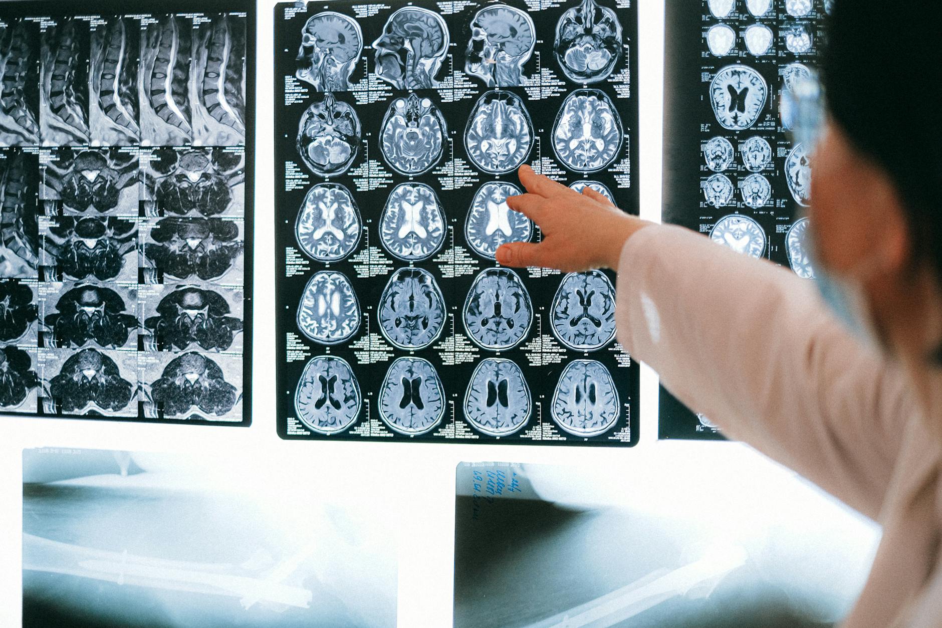
Esaote brings open MRI into the glioma operating room
Italian vendor Esaote presented on May 4, 2026 at the American Association of Neurological Surgeons (AANS) annual meeting in San Antonio the latest update of I-Genius — its first open MRI system designed specifically for intraoperative imaging in glioma surgery. The equipment was developed in partnership with international neurosurgeons to enable multiple MRI acquisitions throughout the procedure without moving the patient off the surgical table. The proposal addresses a well-known clinical problem: during brain tumor resection, distinguishing residual tumor tissue from healthy parenchyma is hard, and surgeons today rely on neuronavigation, electrophysiological mapping and awake craniotomy — useful tools, but they do not deliver three-dimensional evidence of how much tumor has actually been removed.
Following its initial introduction to the neurosurgical community at EANS 2025 in Vienna, I-Genius is now entering a new development phase, with growing international interest and a clear pathway toward U.S. market entry. The system remains under FDA review and is not yet available for sale in the United States, but it is already being discussed as a functional benchmark in neuro-oncology centers.
Why intraoperative MRI is no longer optional
Two decades of studies show that the extent of resection (EOR) is one of the most consistent prognostic factors in high-grade glioma, with direct correlation to overall and progression-free survival. The problem is that intraoperative EOR estimation based on visual inspection and neuronavigation alone is imprecise, especially after the brain shift caused by CSF drainage and tissue removal. Intraoperative MRI systems narrow that error margin by providing fresh imaging during surgery, and multiple published series show higher gross-total resection rates after adopting the technique.
Until now, most iMRI systems on the market run at high field (1.5T or 3T) and require dedicated suites with heavy shielding, intricate workflows and high installation costs. Traditionally, that has restricted access to academic centers. I-Genius takes an alternate path with an open magnet and geometry that lets the patient stay on a single table, reducing operational friction and broadening the set of hospitals able to adopt the technology.
What I-Genius brings to practice
The system is optimized for the surgical environment: the patient stays on the same table throughout the procedure, and the surgeon can trigger multiple MRI acquisitions without repositioning. That shortens procedure time and reduces contamination risk from movement. For neurosurgeon Dr. Roberto Herrera, Chief of Neurosurgery at Clinica Adventista Belgrano in Buenos Aires, "intraoperative MRI systems such as i-Genius change the paradigm of glioma surgery by providing multiple MRI acquisitions directly in the operating room, without disrupting workflow or requiring complex infrastructure, shifting surgery from estimation to objective measurement."
Massimo Olmi, MRI Marketing Director at Esaote, emphasized that the goal is to enhance operating-room efficiency, reduce costs, shorten procedure times and raise the standard of care. The open configuration also offers ergonomic gains for the team — surgeon and anesthesiologist have more maneuvering room than in a closed magnet, which can ease fatigue in long procedures.
Implications for radiology services
Neuro-oncology centers modernizing their neurosurgery fleet should watch I-Genius closely. The combination of open magnet, single table and simplified workflow may make intraoperative MRI feasible at hospitals that currently lack the capital or structure for high-field dedicated systems. The platform model is also coherent with the broader medical imaging trend of valuing workflow and total cost of ownership — themes covered in our interpretation efficiency in radiology guide and the Rochester Regional $20M expansion in oncology and imaging.
For the technology to make it into routine practice, three conditions must be met. First, integration with the hospital PACS so that intraoperative series can be archived, compared and reviewed by the neuroradiology team. Second, joint training of surgical and radiology teams in safe workflows — covering ferromagnetic object policy, MRI-compatible instruments and resuscitation in the magnetic environment. Third, local regulation: regulators must clear the equipment, and the product is still under FDA review, which suggests Latin American availability may follow on a similar window.
Limitations and what to monitor
Open systems carry well-known trade-offs versus closed 1.5T or 3T magnets: lower signal-to-noise ratio, potentially slower gradients and a possibly reduced sequence library. For neurosurgery, however, the metric that matters is the ability to delineate residual tissue, and that depends much more on optimized protocols (FLAIR, DWI, contrast) than on a particular field strength. Clinical results published from early adopters will be decisive in validating the approach.
The surrounding context is favorable. Heavy capital flow into radiology AI indicates that post-processing tools — automated glioma segmentation, residual volume quantification, comparison with the preoperative plan — should reach iMRI routine in the coming years, further amplifying the value of systems like I-Genius. Signals to monitor include FDA clearance, first commercial sites outside Europe, partnerships with surgical equipment vendors and publications with comparative EOR data.


