

Color Imaging in Under One Minute
Scientists from the California Institute of Technology and the University of Southern California have developed a hybrid technique that combines ultrasonography with photoacoustic tomography, producing 3D color images that simultaneously display soft tissue structure and vascular function. The complete exam takes less than one minute and reaches depths of approximately 4 centimeters.
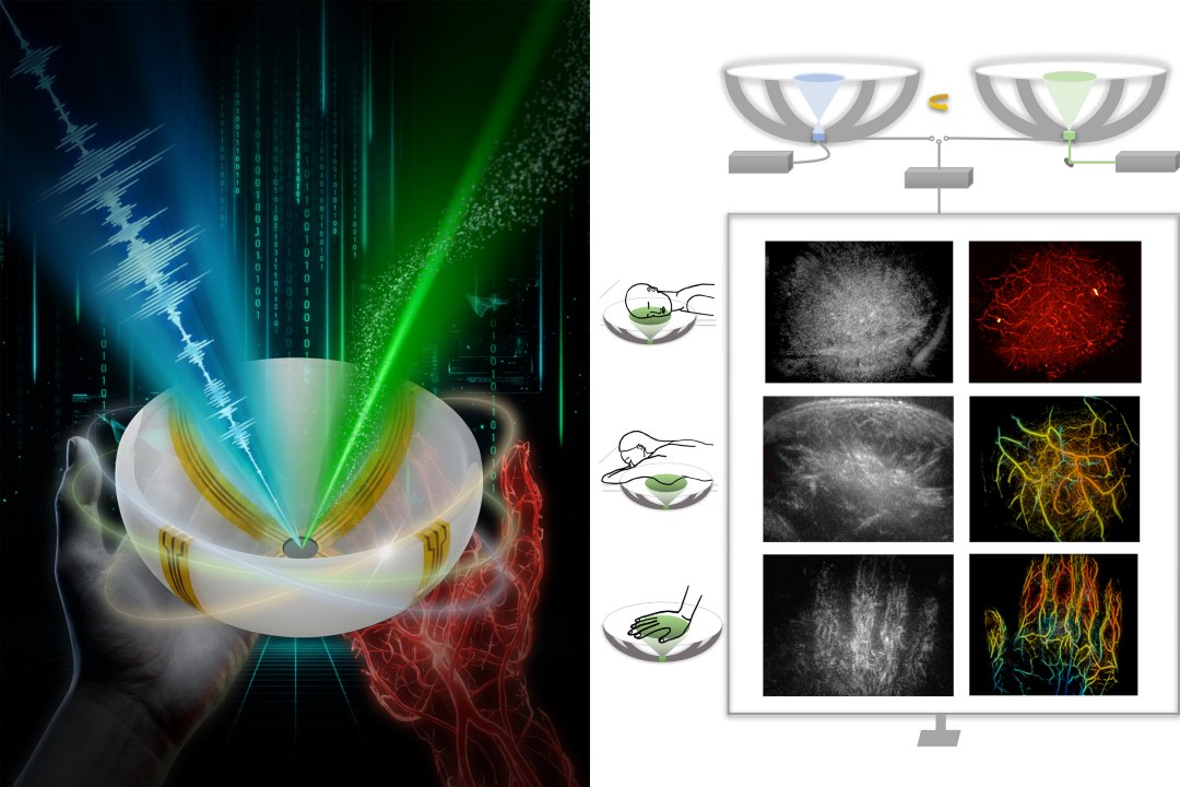
This innovation solves a fundamental limitation of current techniques. Conventional ultrasound offers speed and cost-effectiveness but provides limited field of view in grayscale only. Photoacoustics uses laser to visualize molecules in blood vessels in color but lacks structural detail. The new approach combines the best of both worlds.
How the Technology Works
The team coordinated by Professor Charles Liu used ultrasonic waves to mimic laser excitation, eliminating the need for duplicate equipment. A single transducer emits comprehensive waves through tissues, capturing both anatomical and functional information in a single session.
The study, published in Nature Biomedical Engineering, demonstrates applications across several clinical areas:
- Breast: Tumor detection and visualization with differentiation between healthy and pathological tissue
- Diabetic neuropathy: Monitoring peripheral nerve damage
- Neuroimaging: Brain imaging with vascular detail
- Hemodynamics: Real-time blood flow observation
Impact for Professionals
For professionals working with medical imaging systems, this technology may eventually integrate into existing diagnostic imaging ecosystems. The ability to generate 3D color images with ultrasound equipment represents a significant advance over traditional imaging modalities, especially for screening and point-of-care diagnostics.
Source: Inovação Tecnológica