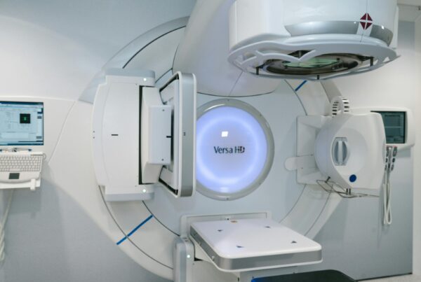

Photon-counting CT (PCCT) has moved out of the research suite and onto fully booked clinical schedules at several US hospitals, and the bar it has to clear has moved with it: sharper images are not enough, the extra information has to change diagnosis, treatment or workflow. That is the thread of a Radiology Today feature in the November/December 2026 issue, written by Keith Loria, who interviewed radiologists at Northwestern Medicine and the University of Wisconsin–Madison and executives at Siemens Healthineers, GE HealthCare and Philips.
Siemens Healthineers received the first FDA clearance for a whole-body photon-counting scanner, NAEOTOM Alpha, in 2021; it is now a line of three models. GE HealthCare’s Photonova Spectra received FDA 510(k) clearance on March 23, 2026, and CE Mark, announced on August 31. Canon launched Ultimion in April, in Japan only, with a cadmium zinc telluride (CZT) detector developed with Redlen Technologies, part of the Canon group since 2021. Philips is still developing the technology through clinical research.
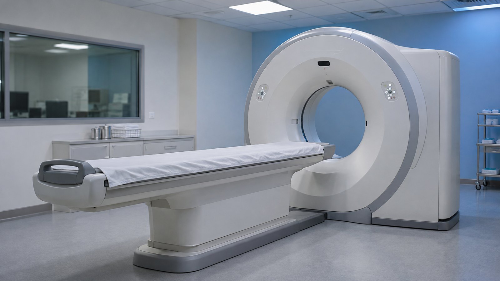
How a photon-counting detector differs from conventional CT
In a conventional energy-integrating detector, X-rays are first turned into light in a scintillator and then into an electrical signal, summing the energy of everything that arrives. A photon-counting detector converts each photon directly into an electrical pulse and measures its energy, sorting events into energy bins (eight, in GE’s silicon design). The result is thinner slices, higher spatial resolution, less electronic noise and spectral information in every exam, with no need to pick a dual-energy protocol in advance.
Those data yield material maps that separate iodine, calcium, fat and water, plus virtual monoenergetic images at different keV levels. Because the spectral information is stored, a radiologist can go back to it when an unexpected question comes up. Lower dose is part of the appeal and builds on a longer trend: CT radiation dose in the US fell 22% over a decade on conventional scanners.
Northwestern: three scanners running at full capacity
Northwestern Medicine in Chicago runs three NAEOTOM scanners, two for inpatients and one at an outpatient center, all at full clinical capacity, according to Amir Borhani, MD, professor of radiology and surgery and medical director of CT at Northwestern Memorial Hospital. Indications are spread across abdominal, cardiac, chest and neuro imaging, and the team tries to route the exams that benefit most to these systems: coronary CT angiography, liver and pancreas, rectal cancer, and pre- and post-transplant studies. Pediatric and pregnant patients are also sent to them, when appropriate, for the lower dose. Matthew Fuld, PhD, director of PCCT at Siemens Healthineers North America, says some US sites perform 100 exams a day and that sites in China exceed 170.
Coronaries, transplants and ablation: where management changes
Cardiac CT is described as one of the clearest use cases. On conventional CT, calcified plaque blooms, making it hard to tell plaque from lumen. Giuseppe Toia, MD, CT modality chief at the University of Wisconsin School of Medicine and Public Health, says that in UW’s evaluation of GE’s silicon-based system, higher contrast-to-noise ratio and lower noise reduced that effect. In at least two patients, he says, conventional CT might have overcalled or undercalled plaque burden, which shifts the choice between medication, watchful waiting and referral to interventional cardiology. Philips coordinates COMBINE-CT, a European consortium of 11 partners now in clinical validation, which includes spectral PCCT follow-up after complex stenting. A photon-counting scanner is also part of the new Hamburg heart and vascular center equipped by Siemens.
In the abdomen, Northwestern has made PCCT its modality of choice after liver transplantation. Borhani says the ultrahigh-resolution mode shows allograft arteries and anastomoses better and that, in an institutional cohort imaged with both PCCT and conventional angiography, the two agreed very closely, with fewer false-positive strictures and fewer unnecessary angiograms. The magazine gives neither the cohort size nor the figures. At UW, after microwave ablation of a liver tumor, Toia’s team combined low-keV monoenergetic images with iodine maps to confirm that zones without iodine held no residual disease, because hemorrhage and protein can raise CT numbers on conventional CT.
Fuld also points to CSF-venous fistulas, a cause of spontaneous intracranial hypotension that can take years to locate. Independent data exist: a retrospective Duke University study in AJNR in 2026, covering 628 CT myelograms in 456 patients, found greater diagnostic certainty with photon-counting CT (OR 1.66; 95% CI 1.17–2.36). Detection was 69.6% versus 61.0% with conventional CT, a difference that was not statistically significant (p = 0.07).
Data volume, protocols and PACS: the price of extra information
Toia estimates that a PCCT study generates about 20 times as much data as a conventional CT exam; GE says Photonova’s GPU-accelerated reconstruction is designed for up to 50 times more data than its own flagship conventional scanner. Thin slices and multiple spectral series weigh on reconstruction, networks, storage and reading. The answer, Toia says, is not to switch on every feature for every patient: a pancreatic mass justifies ultrahigh resolution, nonspecific abdominal pain probably does not. “You have to balance the clinical value with the technological advantage,” he says.
Standardization is under way. A Society of Abdominal Radiology consensus published in AJR in December 2025, with Toia and Borhani among the authors, brought together nine radiologists from nine institutions and agreed on 20 abdominal protocol features, such as 70-keV monoenergetic images for primary reading of portal venous and pancreas PCCT. The survey also covered which spectral reconstructions go to PACS and how the spectral dataset is archived. For any department weighing the technology, that is the decision to plan early: what goes to PACS, for how long, and with what network capacity and PACS integration and disaster recovery plan.
Dose calls for the same judgment: larger patients, motion or arms that cannot be raised may still yield diagnostic images without a dose increase, while routine exams leave room to lower it. On cost, Fuld says the decision depends on patient mix, throughput and whether clinicians are ready to redesign protocols instead of running the scanner like a conventional system.
Limitations and what comes next
The feature is a collection of interviews, not a study. Several voices are vendors, and Toia said in a March GE press release that he had been involved in developing and testing the Deep Silicon detector. The examples of changed management are single cases or institutional series without numbers in the article. Toia expects cardiac imaging to move fast and abdominal imaging to need longer studies tying clearer lesions to staging, treatment and outcomes. “Yes, we are getting better images, and that makes radiologists more confident in what they are looking at,” he says. “But does that translate to a difference in care? There needs to be more data collection on that.”
Further out, researchers at the University of Victoria and Redlen are using the Canadian Light Source synchrotron to study the few-atoms-thick oxide layer that controls electron extraction from the CZT detector, and are testing a sulfide as an alternative. Another goal is to tell two or more contrast agents apart in one scan; Fuld says current systems can already record the data, but new agents still have to be developed and approved.
Source: Radiology Today — Sharper Imaging. Studies cited: Christensen et al., AJNR 2026; Dane et al., AJR 2025.