
Women with invasive breast cancer whose preoperative breast ultrasound showed a high glandular tissue component were about twice as likely to develop a new cancer in the same or the opposite breast in the years after surgery. The finding comes from an 8,886-patient study at two tertiary centers, one in South Korea and one in China, published in the November issue of Radiology. The same marker showed no relationship with regional or distant recurrence.
This is an association seen in retrospective cohorts, not a validated tool for changing follow-up. What makes it interesting is that the rating is made on the ultrasound exam itself, with no contrast, no radiation and no added cost, and that it has just entered the BI-RADS ultrasound lexicon.
How the Radiology study assessed 8,886 patients
The paper is led by Tingting Deng of the Department of Ultrasound at Sun Yat-sen University Cancer Center in Guangzhou and Woo Kyung Moon of Seoul National University Hospital, with 15 other co-authors. It included consecutive women who had surgery for invasive breast cancer from 2013 to 2018 at the Korean institution (5,167 patients, mean age 52.4 years) and from 2013 to 2019 at the Chinese institution (3,719 patients, mean age 49.2 years).
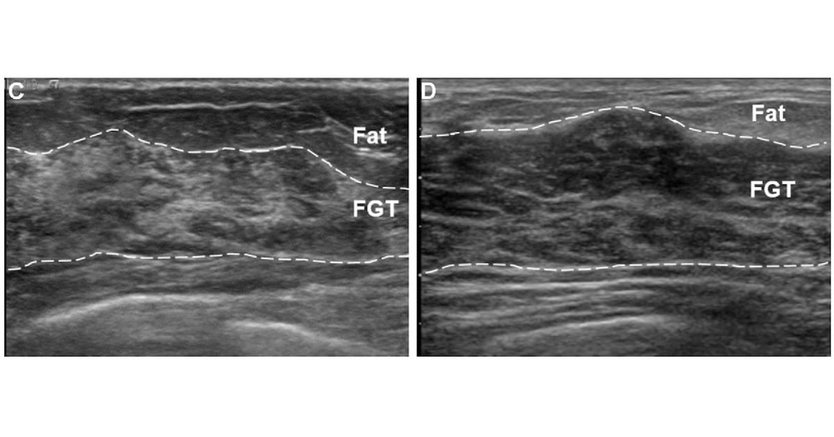
The glandular tissue component (GTC) was graded on preoperative bilateral ultrasound images and grouped as low (minimal or mild) or high (moderate or marked). The two sites did this differently: in Korea, the category was recorded prospectively in routine reports; in China, it was assigned later from archived images. The analysis used Fine-Gray competing-risk models adjusted for clinicopathologic variables, so the results are subdistribution hazard ratios. Median time from surgery to recurrence or last follow-up was 6.8 years in Korea and 8.0 years in China.
Higher risk of new breast cancer, not of metastasis
An ipsilateral or contralateral breast cancer occurred in 145 patients in Korea (2.8%) and 85 in China (2.3%). Regional or distant recurrence occurred in 233 (4.5%) and 520 patients (14.0%), respectively. In multivariable analysis, high GTC was associated with more ipsilateral or contralateral cancer at both sites, with hazard ratios of 1.93 (P < .001) and 2.02 (P = .002). For regional or distant recurrence, the figures were 0.89 (P = .46) and 1.20 (P = .06), which the authors describe as no evidence of an association. At the Chinese center that result sat close to the significance threshold, so a flat “no association” reads the data more firmly than they allow.
The cumulative incidence curves also separated when the two events were looked at on their own: ipsilateral cancer, in an analysis limited to patients treated with breast-conserving surgery, and contralateral cancer. Keep the scale in mind. Fewer than 3% of patients developed a new breast cancer during follow-up, so doubling a small risk still leaves a modest absolute risk, and the abstract reports neither absolute rates by group nor confidence intervals.
“If this marker is validated in future studies, patients with high GTC at preoperative US may potentially benefit from tailored surveillance strategies, including supplemental imaging approaches,” Deng and colleagues wrote, as quoted by Diagnostic Imaging.
Density, glandular tissue and background enhancement: what GTC measures
Mammographic density is an established risk factor, but mammography cannot separate the two parts of fibroglandular tissue: ducts and lobules look just as white as fibrous stroma. Ultrasound can tell them apart. Terminal duct lobular units, the glandular component, appear relatively hypoechoic, while interlobular fibrous tissue is hyperechoic. GTC estimates the glandular share of the fibroglandular tissue as minimal (under 25%), mild (25%–49%), moderate (50%–74%) or marked (75% or more).
The proposed biology is lobular involution. Lobules regress with age, and incomplete involution has been linked to higher cancer risk in studies of benign biopsies. In 2021 the Seoul group reported in Radiology on 8,483 women with dense breasts and negative mammograms: moderate or marked GTC at screening ultrasound was associated with a 1.5-fold higher cancer risk (95% CI: 1.05, 2.1) after adjustment for age and density, and the category tracked the degree of involution seen at biopsy.
On MRI, the amount of fibroglandular tissue and background parenchymal enhancement (BPE) play a similar role. A 2023 Radiology review places BPE and GTC side by side as markers that add to density, each in its own modality. The BI-RADS v2025 edition added GTC to the ultrasound tissue composition item, next to background echotexture, with the same four categories, according to the ACR’s lexicon summary form.
What it could change in post-treatment surveillance
New ipsilateral and contralateral cancers are exactly what breast imaging surveillance tries to catch early. Annual mammography is the usual backbone, and supplemental imaging is added for subgroups at a cost in false positives. In a prospective study in JAMA Oncology in 2017 of 754 women aged 50 or younger treated with breast conservation, adding MRI raised detection from 4.4 to 8.2 cancers per 1,000 exams and adding ultrasound raised it to 6.8 per 1,000, while specificity fell from 96% to 87% and 88%. The same trade-off shows up in screening, where a recent study questioned supplemental ultrasound after a negative DBT because of its false-positive burden.
A marker such as GTC could help decide who gets the extra exam, and other routes to risk stratification are being tried, such as the MRI-based AI for breast recurrence risk the FDA recently cleared. In services where breast ultrasound is already part of staging or follow-up, recording the category needs no new exam. Standardization is the weak point. Groups are testing AI and automated breast ultrasound (ABUS) for GTC grading, while products such as DeepHealth’s breast ultrasound AI still target detection and reporting, not risk.
Limitations and what is still missing
The authors themselves point to the retrospective design, possible differences in how the two institutions rated GTC, and the composite endpoint. Combining ipsilateral events, which may be the original tumor returning or a new primary, with contralateral cancer mixes outcomes with different biology and management. The rating is visual and subjective: in a 2017 study with 11 radiologists, interobserver agreement was moderate, with kappa values of 0.45 for the four categories and 0.48 for the two-group version. The abstract also does not detail which treatment variables entered the adjustment.
The cohort included only Asian women, from populations where dense breasts are common and ultrasound plays a central role. Even within that group, breast cancer risk varies widely across subgroups, and there are no GTC data from Latin American or other populations. The next step is prospective validation in cohorts of other backgrounds, ideally with automated measurement, to see whether the marker adds information to the risk models already used in follow-up.
Source: Diagnostic Imaging — Breast Ultrasound Study: High Glandular Tissue Component May Double Recurrence Risk for Invasive Breast Cancer. Original paper: Deng T, Moon WK et al. Glandular Tissue Component at Preoperative Breast US and Recurrence Risk in Invasive Breast Cancer. Radiology 2026;321(2):e260681.