Meta will give three Ohio State University research teams access to its Muse AI models, along with grant money, for projects in radiology and clinical AI: drafting chest X-ray reports, predicting heart failure risk from routine CT scans, and building an agent system to support Alzheimer’s diagnosis. Meta and the Ohio State Wexner Medical Center announced the work on October 28 as part of a program spanning five US universities.
The models come from Meta Superintelligence Labs (MSL), the company’s AI research and development division. According to Meta, the universities will run the models inside their own environments and keep control of all patient and clinical data, which will not be shared with or processed by Meta. No results exist yet; what was announced are research plans.
What Meta announced, and how the money is split
Besides Ohio State, the program covers Caltech, Louisiana State University, Indiana University and UTHealth Houston. Support differs by project and can include model access, collaboration with Meta researchers and, for some projects, funding. Meta’s newsroom post puts the total at USD 2 million across all five schools. Radiology Business described the Ohio State projects as funded by a grant of roughly USD 2 million; Ohio State’s own release gives no figure, and per-project amounts have not been disclosed.
The Muse family includes closed models offered through an API and open-weight models that can be downloaded and run on local hardware, such as the 816-billion-parameter Muse Spark Open and the 30-billion-parameter Muse Glimmer, which fits on a single consumer GPU once quantized. Neither announcement says which model each Ohio State team will use.
Chest X-ray drafts, CT-based cardiac risk and Alzheimer’s agents
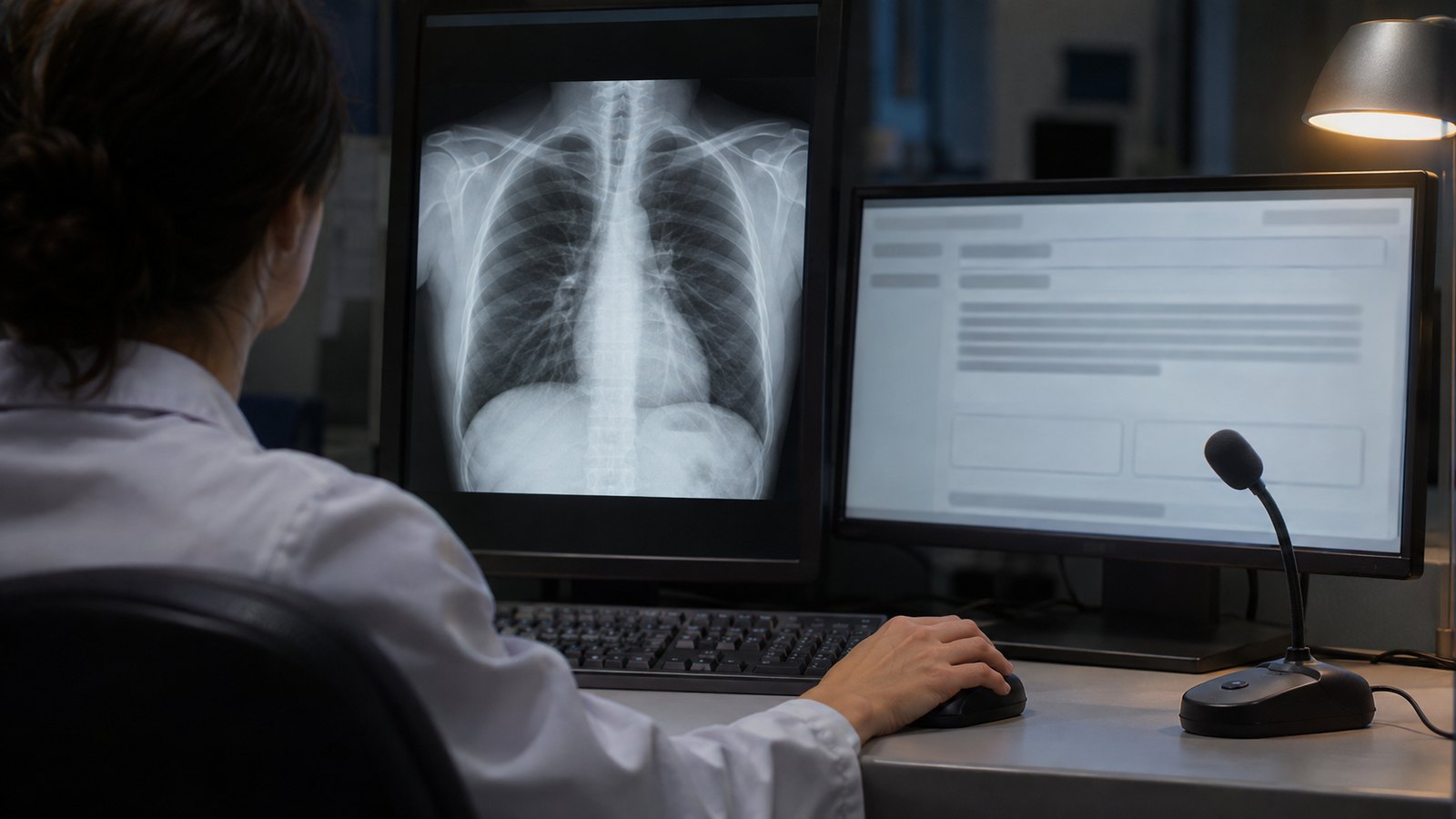
Drafting chest X-ray reports. The project closest to daily radiology practice is led by Dufan Wu, PhD, associate professor of radiology, with radiologists Chiemezie Amadi and Paul Lewis. Meta says Ohio State’s thoracic radiologists read about 200 chest X-rays a day, and some institutions reach 1,000. Most published report-generation models look only at the image, whereas a radiologist also weighs the reason for the exam, the patient’s history and prior studies. The team wants the draft to use all of that.
Two further pieces get less attention. One corrects, statistically and against a small fixed set of human-annotated reports, the bias that creeps in when one language model grades another model’s report. The other, run with psychologist Peter Kvam, PhD, is a reader study in a simulated clinical setting that separates under-reliance (rejecting correct AI content) from over-reliance (accepting incorrect content) and links each behavior to the kinds of errors the model makes.
Heart failure with preserved ejection fraction. HFpEF accounts for roughly half of heart failure cases and is often recognized late because its symptoms overlap with obesity and other common conditions. The idea is to reuse abdominal CT scans ordered for unrelated reasons, which already capture body fat distribution, liver fat, muscle composition and vascular calcification, and combine them with longitudinal records and ECG data when available. The model should return a risk estimate with the findings behind it and cope with missing inputs. Yuan Xue, PhD, assistant professor of biomedical informatics, leads with cardiologist Yuchi Han, MD, and AI researcher Xia Ning, PhD.
Clinician-guided agents for Alzheimer’s diagnosis. Ning, Xue and cognitive neurologist Douglas Scharre, MD, are building a clinician-in-the-loop multi-agent system on open-weight Muse models deployed locally. The agents should reconstruct a patient’s cognitive history, flag diagnostic uncertainty and recommend the next most informative assessment, drawing on records from Ohio State’s Memory Disorders Clinic, which handles more than 5,600 patient visits a year.
“We also seek to understand how physicians can optimally interact with AI and how these systems can be made more reliable and useful in clinical practice,” said Orlando Simonetti, vice chair of research in Ohio State’s Department of Radiology.
Automation bias and opportunistic CT: the technical background
The reader study addresses a well-documented problem. In a 2023 Radiology experiment, Dratsch and colleagues had 27 radiologists assign BI-RADS categories to mammograms with help from a purported AI that sometimes suggested the wrong category. When the suggestion was wrong, accuracy fell from 79.7% to 19.8% among inexperienced readers and from 82.3% to 45.5% among very experienced ones. Real-world errors look similar, as in the case of an AI that flagged a pneumothorax that was not there after pacemaker implantation.
Ohio State’s design puts clinical context into the input and studies reader behavior before deployment. General-purpose models used without that groundwork can fail badly, as a recent test showed when ChatGPT-5 missed half of distal radius fractures. Open models have reached radiologist-level results in narrower tasks, such as the open-source AI evaluated on prostate MRI in SCARLET-1, but free-text report drafting is a broader problem.
Opportunistic CT has a track record too. In 2020, Pickhardt and colleagues ran automated algorithms on abdominal CT scans from 9,223 asymptomatic adults and followed them for a median of 8.8 years. In The Lancet Digital Health, automated aortic calcification predicted five-year mortality with an AUC of 0.743, compared with 0.688 for the Framingham score. Ohio State builds on those measures but narrows the target to HFpEF and adds records, ECG and a written rationale.
Why local, open-weight deployment matters for hospitals
The most transferable part of the announcement is the deployment model. With open weights, inference runs on hospital servers and data stay inside the institution. For readers in Brazil, the LGPD treats health information as sensitive personal data (Art. 11) and sets separate rules for international transfers (Art. 33). Running a model locally avoids sending images and reports abroad, but it does not remove the need for a legal basis, access controls and governance over secondary use of records. And “local” depends on size: a quantized 30-billion-parameter model fits on a 24 GB to 32 GB GPU, while the 816-billion-parameter model needs hundreds of gigabytes of memory.
How these models write reports in Portuguese or Spanish, with local terminology, is untested. Once a tool like this drafts reports or produces risk scores in routine care, it becomes software as a medical device, regulated in Brazil under Anvisa’s RDC 657/2022 and by the FDA in the United States; the current projects are research. The workforce question also remains contested, as the Neiman Institute’s rebuttal of the claim that AI replaces radiologists shows.
Limitations and what to watch next
None of the three projects has a published protocol, sample size, predefined endpoints or timeline. Meta evaluated the Muse models on agent, coding and reasoning benchmarks, not clinical tasks, and none of them is a cleared medical device. Meta says faculty designed the studies; even so, it is worth watching whether results compare Muse with other open models and whether they appear in peer-reviewed journals. As discussed at SIIM-CAIMI 2026, imaging AI has moved from evaluation to implementation, and calibrated user trust, which Ohio State’s reader study aims to measure, sits at the center of that shift.
Source: Radiology Business — Meta to fund radiology AI research at Ohio State. Primary sources: Ohio State Wexner Medical Center and Meta Research.