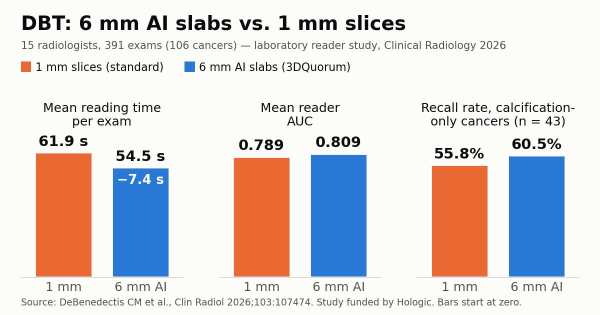
Reading digital breast tomosynthesis (DBT) as 6 mm AI-generated slabs instead of standard 1 mm slices cut mean reading time from 61.9 to 54.5 seconds per exam with no loss of diagnostic accuracy, according to a multireader study published in Clinical Radiology. Fifteen radiologists read the same 391 exams both ways, using Hologic’s 3DQuorum software.
That is 7.4 seconds per case. The authors report it as a 13% reduction, a figure that comes from the study’s mixed-regression model; dividing the two published means gives about 12%. Either way, the saving is small per exam and only matters once it is multiplied across a screening worklist.
How the 15-reader study was set up
The first author is Carolynn M. DeBenedectis, a professor of radiology at UMass Chan Medical School in Worcester, Massachusetts. The other five authors work in Hologic’s research and development department, and Hologic funded the study. Exams were collected retrospectively from women imaged in the United States between January 2016 and April 2017 and selected in a stratified, randomized way: 160 negatives, 27 recalls, 98 biopsy-proven benign lesions and 106 biopsy-proven cancers.
The design was fully crossed, so every reader interpreted every case in both modes. The primary hypothesis was non-inferiority of the area under the ROC curve (AUC). Mean AUC was 0.809 with the slabs and 0.789 with 1 mm slices, a difference of +0.020 (95% CI 0.002 to 0.037). The interval sits entirely above zero, but the gap is small and the trial was built to show the new display is not worse, not that it is better.
The second endpoint targeted the weak spot of any thick reconstruction: the 43 cancers that presented as calcifications only. Readers recalled 60.5% of them with slabs and 55.8% with thin slices (difference +0.047; 95% CI 0.005 to 0.088). The authors call this result “particularly important because calcifications may represent DCIS or early invasive disease and may be vulnerable to reduced conspicuity with thicker slab reconstructions.” The abstract also reports no evidence that the effect differed by breast density.
MIP slabs versus AI-weighted slabs
Tomosynthesis eased the tissue-overlap problem of 2D mammography but made reading slower. In a 2014 Massachusetts General Hospital study in Radiology, combined tomosynthesis and mammography took a mean 2.8 minutes per screening exam versus 1.9 minutes for digital mammography alone, or 47% longer. By Hologic’s own arithmetic, a 60 mm compressed breast produces about 240 one-millimeter slices across a four-view screening set.
Collapsing thin slices into thicker ones is the obvious fix, but the method matters. Simple summing or averaging dilutes small structures such as microcalcifications across the slab. Maximum intensity projection (MIP) keeps the brightest pixel through the slab, which makes dense calcifications pop but reintroduces tissue overlap and, according to the authors, can hide small clusters of amorphous calcifications and subtle architectural distortion.
3DQuorum takes a different route. According to Hologic’s technical white paper, a CAD-like algorithm first scans the 1 mm slices for bright spots that could be calcifications, radiating lines suggesting spiculation or distortion, and rounded dense objects, and logs where each one sits. It then merges six 1 mm slices into each 6 mm SmartSlice, giving extra weight to the flagged regions. Neighboring SmartSlices overlap by 3 mm, which cuts the image count to roughly one third. The FDA approved the technology in November 2019.
An FDA-era trial, now peer reviewed, plus real-world data
One piece of context is missing from the Diagnostic Imaging coverage. Hologic’s 2019 white paper describes a reader study with the same design and the same figures (391 cases, 15 readers, 61.9 and 54.5 seconds, “about a 13% reduction”) that was presented to the FDA. The 2026 paper appears to be the peer-reviewed version of that regulatory trial. The same document says the slab arm was read alongside the company’s higher-resolution synthesized 2D image (Intelligent 2D, 70 µm) while the standard arm used C-View, so not all of the difference can be pinned on slab thickness alone.
Whether detection holds up in routine screening, which a reader study cannot answer, now has a first answer. A review of 119,662 screening DBT exams at Massachusetts General Hospital, published in Radiology in 2026 and covered in our roundup of the breast imaging studies that defined 2026, compared periods before and after the switch to 6 mm slabs with 3 mm overlap. Cancer detection (5.8 and 6.5 per 1,000) and sensitivity were non-inferior, the abnormal interpretation rate fell from 6.2% to 5.8%, and specificity rose from 94.4% to 94.9%. That study did not measure reading time.
What it means for breast imaging practices
Breast AI tools solve different problems. This one is neither a lesion detector nor a risk model, a distinction covered in Breast AI: detection and risk models aren’t interchangeable; it changes how the image is displayed. The goal is throughput, the same pressure behind Stockholm’s move to replace the second mammography reader with AI, reached by a different route.
There are side effects beyond reading time. Hologic estimates files about 60% smaller, which eases PACS storage and network load, but only once the 1 mm slices stop being archived; during a transition the vendor lets sites send both series, which increases volume. SmartSlices are generated on the acquisition workstation of Hologic systems, so the option is tied to one vendor. Practices weighing the switch outside the United States should confirm local regulatory status, and any site should compare recall rate, positive predictive value and cancer detection before and after, the kind of audit that BI-RADS v2025 puts front and center.
Limitations: an enriched case set and industry funding
The main limitation, acknowledged by the authors, is enrichment: 106 of 391 cases (27%) were cancers, while screening detects around 6 per 1,000 exams, as in the MGH series. Readers who meet a cancer every fourth case behave differently, and time spent on positive cases does not distribute the way it does on a real worklist. A laboratory read also lacks prior exams, interruptions and clinical pressure.
The breast density analysis was exploratory and underpowered. Finding no difference in dense breasts does not show that the benefit holds there, and dense breasts are already where DBT struggles, as discussed in is screening ultrasound still needed in the DBT era? Industry authorship, images from 2016 and 2017 and the different synthesized 2D images in each arm also weigh on interpretation.
Hologic’s long-standing claim of one hour saved per eight-hour reading day is an extrapolation of the 13% figure and assumes the whole shift is spent scrolling slices. The useful next step is a prospective screening-population study that tracks reading time, recall and DCIS detection together.
Source: Diagnostic Imaging — Mammography Study Reveals Nearly 13 Percent Reduction in Reading Time with AI-Powered Slab Reconstruction for DBT. Primary paper: DeBenedectis CM et al., Clinical Radiology, 2026.