
What MRI finds in scapular pain also shows up in people without it
MRI findings in patients with pain around the shoulder blade overlap with findings that occur in
the general ageing population, according to research published on October 16, 2026 in
Interventional Pain Medicine. A team led by Chetan Potu, of Stanford University in
California, identified C5-6 pathology and supraspinatus tendinopathy as the most prevalent imaging
findings — precisely the two that the literature already links to age.
The authors’ conclusion is a warning about interpretation, not a dismissal of the exam:
“Incidental MRI findings in the spine and shoulder increase with age, and therefore MRI findings in
patients with scapular pain should be cautiously interpreted within appropriate clinical
context.”
Why scapular pain is a hard imaging problem
Pain in the scapular region can arise from several sources: the cervical nerve root, the
glenohumeral joint, the rotator cuff, the scapulothoracic articulation, periscapular musculature,
the thoracic spine or referred visceral pain. Correctly identifying the source matters because it
determines the interventional strategy — a nerve root block, a subacromial injection, a facet block
or a muscular approach are distinct procedures with distinct targets.
In practice, clinical history and physical examination drive most decisions, and MRI is called on
when isolated scapular pain fails to improve over time. What was missing, the authors note, was data
on when clinicians decide to order MRI, which region they order and which abnormalities appear most
often. The study was designed to fill that gap.
Design and numbers
The study included 503 patients seen at a single centre between 2015 and 2025 who presented to
orthopaedic clinics with scapular pain. Of those, 284 underwent at least one MRI exam. The
distribution by region reveals the ordering pattern: cervical spine in 203 cases, shoulder in 106,
thoracic spine in 76 and the scapula itself in only 22.
Because the total exceeds the number of patients, some of them had more than one exam. The
authors state the hypothesis honestly: “While the reason for additional imaging was not assessed in
the present study, it is possible that the imaging findings from the first MRI did not sufficiently
explain the etiology of the scapular pain, or that treatment of that anatomic region did not improve
the scapular pain.” In other words, the second exam tends to follow a first one that failed to
answer the clinical question.
Finding prevalence and the specificity problem
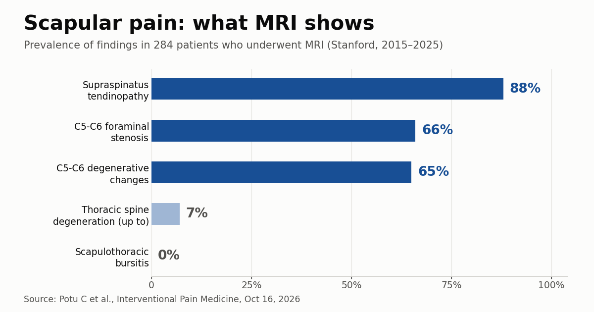
The most frequent findings were supraspinatus tendinopathy (88%), C5-6 foraminal stenosis (66%)
and C5-6 degenerative changes (65%). All showed significant associations with age (p < 0.01 for
all) but not with sex — a result that reinforces earlier studies on the rising prevalence of
supraspinatus tendinopathy and rotator cuff disease over a lifetime.
At the other end, the least common findings were scapulothoracic bursitis (0%) and thoracic spine
degenerative changes (0% to 7%). Notably, the team observed no association between the laterality of
the scapular pain and the laterality of cervicothoracic spine findings: pain on one side did not
predict a finding on the same side.
Together, that draws the problem clearly. A finding present in 88% of a population selected for
pain, strongly age-dependent and uncorrelated with the symptomatic side, has low specificity for
explaining the complaint. It can be present and still be irrelevant.
Practical implications for reporting and referral
For the radiologist, the lesson concerns wording. Describing supraspinatus tendinopathy or C5-6
foraminal stenosis without qualifying how prevalent those changes are in the patient’s age group
hands the clinician a datum that looks explanatory and often is not. A report that records the
finding and adds that it is common in asymptomatic people of the same age is more useful — and
reduces the chance that the exam becomes justification for a procedure aimed at the wrong
target.
For whoever organises the workflow, 284 exams across 503 patients, with multiple regions ordered,
points to potential waste. It is worth assessing whether a clearer indication protocol would reduce
the chaining of exams — the same efficiency discussion we raised when covering
the cost of MRI repeats.
And the theme connects directly to the broader debate about
how to handle incidental findings,
where the presence of an abnormality is not the same as causation.
Limitations and what comes next
The design is retrospective, single-centre, and spans a decade over which MRI protocols and
reporting criteria changed. There was no control group of patients without scapular pain undergoing
the same exams, which would be the direct test of the overlap hypothesis; the authors rely on
comparison with the literature. Outcomes were not assessed either: it is unknown whether patients
who had MRI fared better.
The researchers themselves call for prospective studies with standardised ordering and
interpretation, able to determine the clinical utility of imaging in this setting. It is the kind of
question musculoskeletal radiology has been facing in other joints — including with quantitative
tools, as we covered in
radiomics for grading osteoarthritis
— and one that will only grow in relevance as imaging becomes more accessible and the risk of
incidental findings rises in step.
Source: AuntMinnie — MRI shows features of scapular pain in patients