

An open-source AI system for prostate MRI matched radiologists in detecting clinically significant prostate cancer across three external cohorts, one diagnostic and two from population-based screening. That is the main finding of the SCARLET-1 study, published in Radiology by Anindo Saha and colleagues at Radboud University Medical Center in Nijmegen, the Netherlands, with areas under the curve (AUC) of 0.84 to 0.89 and agreement with the reference diagnosis 13 to 16 percentage points above that estimated for an average radiologist.
What SCARLET-1 tested
SCARLET-1 stands for Scaling Artificial Intelligence for Prostate Cancer Detection on MRI to Global, Multiethnic Populations. It is a retrospective, confirmatory validation study with a statistical analysis plan registered in advance on arXiv and reporting aligned with STARD-AI and TRIPOD+AI. The investigators pooled 22,445 prostate MRI examinations from 21,252 men with suspected prostate cancer, acquired between January 2012 and October 2024 in 46 cities across 22 countries, including sites in Vietnam, Kenya, China and the United States.
The algorithm under test is PI-CAI-2B, a computationally lighter successor to the system that came out of the international PI-CAI (Prostate Imaging: Cancer AI) challenge. It was trained in a single run on 18,471 examinations from 26 cities in 14 countries, with no subsequent retraining or redesign, and its source code and model weights are public on GitHub. Scans came from 38 different 1.5 T and 3 T scanner models from five vendors.
The part that matters most for clinical practice is the external test, which covered 1,974 patients from 20 cities in 12 countries, drawn from three previously published prospective trials:
- PRIME (n = 476): diagnostic setting, men referred with clinical suspicion, 30% prevalence of significant cancer;
- STHLM3-MRI (n = 1,108): organized population-based screening in Sweden, 17% prevalence;
- IP1-PROSTAGRAM (n = 390): population-based screening in the United Kingdom, 4% prevalence.
One clarification: some coverage described the study as involving “2k patients.” That figure refers only to the external test cohort (1,974); the study as a whole included more than 21,000 men.
The headline numbers
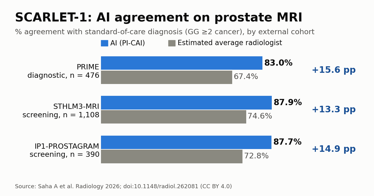
The primary outcome was agreement between the AI assessment and the standard-of-care diagnosis of Gleason grade group (GG) ≥2 cancer. The AI agreed in 83.0% of cases in PRIME, 87.9% in STHLM3-MRI and 87.7% in IP1-PROSTAGRAM. Expected agreement for an average radiologist was 67.4%, 74.6% and 72.8%, respectively, an advantage of roughly 15.5, 13.3 and 14.9 percentage points for the algorithm. Prespecified criteria for diagnostic interchangeability were met in all three datasets.
External AUCs were 0.86 in PRIME, 0.89 in STHLM3-MRI and 0.84 in IP1-PROSTAGRAM; the last one carries a wide confidence interval (0.70 to 0.95), reflecting low prevalence. Decision curve analysis showed positive net benefit both at cancer-averse thresholds (10% to 30%) and at biopsy-averse thresholds (above 30%).
An important methodological caveat: the “average radiologist” comparator is not a local re-read. It is a statistical estimate derived from the PI-CAI observer study, in which 62 radiologists from 45 centers in 20 countries read 400 examinations, adjusted for the prevalence of each cohort. The authors themselves acknowledge that this benchmark may be optimistic relative to real-world variability.
Biopsies: the effect depends on the population
In the subset of patients with histologic verification, the authors compared the AI with the original radiology readings (PI-RADS ≥4) at a high-specificity operating point (88% to 90%) with 73% to 76% sensitivity. In STHLM3-MRI, the AI detected 26.1% more high-grade cancers (145 vs 115) but generated more than twice as many unnecessary biopsies (47 vs 21). In IP1-PROSTAGRAM, the result went the other way: 19.2% fewer unnecessary biopsies (21 vs 26) while detecting the same number of GG ≥2 cancers (11 vs 11).
In PRIME, at a high-sensitivity operating point (90% sensitivity, 58% specificity), the AI generated 34.2% fewer unnecessary biopsies but detected 9.1% fewer cancers than the historical readings. The authors’ message is that the operating threshold is a design choice that must fit the clinical workflow, not a fixed property of the algorithm.
Technical context: why external validation matters
Pre-biopsy multiparametric MRI is now the international standard of care, and the case for MRI-based screening gained weight when Cochrane shifted its position on prostate cancer screening. The bottleneck is interpretation: PI-RADS reading varies widely between radiologists and relies on genitourinary expertise concentrated in high-volume centers. Even in the United States, pre-biopsy MRI is used in only about a third of cases, and MRI is also gaining ground in local staging of prostate cancer.
SCARLET-1 also exposes the limits of generalization. Performance was stable across age bands, ethnic groups, tumor location and risk strata (mean AUC difference of 0.02), but varied considerably across the 25 sites: 28% of them had an AUC below 0.80, while 32% reached 0.90 or higher. Image quality mattered a great deal, with an AUC of 0.78 in examinations rated PI-QUAL 1 (inadequate) versus 0.88 in PI-QUAL 3 (optimal). For anyone running an MRI service, this is a reminder that acquisition protocols, patient preparation and quality control remain decisive, with or without AI.
Practical implications
The authors’ central argument is that AI can extend prostate MRI to places where subspecialty readers are scarce, a situation common across much of Latin America, Africa and Asia, and in rural areas of high-income countries. An open model trained on multiethnic data and backed by published external validation is a useful starting point for research groups and services that want to test the tool on their own data. It does not, however, replace regulatory clearance for clinical use or local validation.
The Radboud group explicitly recommends site-level validation and continuous performance monitoring, because shifts in case mix, referral patterns or equipment can erode accuracy without being obvious. They suggest recalibrating or moving the operating point when differences stem from prevalence, and retraining or fine-tuning when performance drops across all thresholds. The discussion connects with other fronts in prostate diagnosis that we have covered, such as PSMA PET/CT-guided biopsy in men with negative MRI.
Limitations and next steps
The study is retrospective and combines consecutive patients in the external test sets with convenience samples in training. Several subset analyses involve small samples. The global operating point used for the interchangeability test was prespecified for methodological consistency and was not intended for clinical use. Moreover, most men with a negative MRI did not undergo biopsy, which prevents a full comparison of false-negative rates between AI and radiologists and leaves the results exposed to partial verification bias.
In short, SCARLET-1 supports the claim that AI performs on par with standard-of-care reading, not that it decisively outperforms it. Ongoing prospective trials should clarify how to use continuous risk scores, combined with clinical data and patient preferences, rather than a binary “biopsy or not” output.
Source: Saha A, Bosma JS, Twilt JJ et al. Artificial Intelligence for Prostate Cancer Detection and Screening on MRI in Global Populations: The SCARLET-1 Study. Radiology 2026;321(1):e262081. doi:10.1148/radiol.262081; Diagnostic Imaging; The Imaging Wire.



