

A CT bone reading software that “unfolds” the ribs and displays them side by side in a single plane helped two radiologists find more rib metastases in women with breast cancer: 22.5% and 8.3% more lesions, respectively, than conventional axial reading. The retrospective, single-center study was led by Derya Karabulut and Aykut Alkan of the Department of Radiology at Trakya University in Edirne, Turkey, and published online on October 12 in Academic Radiology. Beyond finding more lesions, the tool lowered the minimum lesion size detected and cut reading time roughly in half.
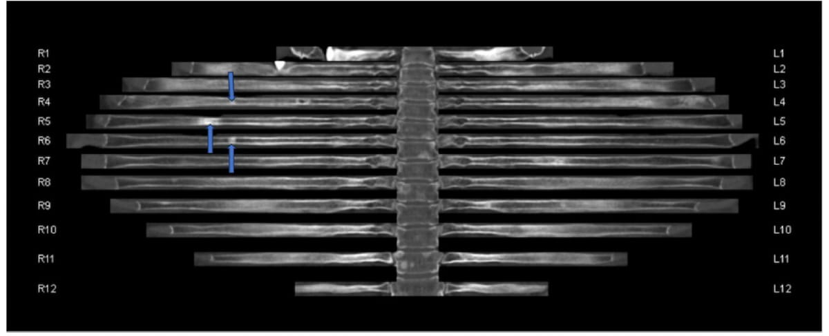
What the study evaluated
The authors reviewed 110 patients with breast cancer and bone metastases (median age 56) who underwent thoracic CT and PET/CT between January 2022 and January 2026; 82 of them had rib lesions. The reference standard combined PET/CT with available longitudinal follow-up imaging for each suspected lesion. Two radiologists independently read the same exams in two sessions at least two weeks apart: one with conventional CT and one with bone reading post-processing (syngo.via CT Bone Reading, version VB80, Siemens Healthineers).
In the lesion-based analysis, conventional reading allowed reader 1 to identify 510 lesions (81.6% of the reference total) and reader 2 to identify 577 (92.3%). With the tool, reader 1 found 115 additional lesions and reader 2 found 48 more. Those are the 22.5% and 8.3% gains quoted by Diagnostic Imaging; the proportion is relative to what each reader had already found, not to the total lesion count. Based on the abstract’s percentages, the reference standard included roughly 625 rib lesions.
Three findings stand out:
- Where the extra lesions were: about half of them (47.8% and 50%) sat in the mid-rib, mainly at levels 5 to 8.
- Minimum size: the median smallest detected lesion fell from 8 mm to 5 mm for reader 1 and from 7 mm to 5 mm for reader 2 (P < 0.001).
- Reading time: from 129.22 s to 64.94 s for reader 1 and from 122.99 s to 64.44 s for reader 2, according to the abstract indexed in PubMed. The Diagnostic Imaging report gives “64.44 to 18.71 s” for the second reader, but 18.71 s is the standard deviation; the actual reduction was close to 48%.
Inter-reader agreement with the tool was excellent (ICC ≥ 0.965; kappa ≥ 0.947). The authors attribute the time savings mainly to automatic labeling of each rib level and to seeing every arc in one plane. According to the abstract, the less experienced reader gained the most.
Why the mid-rib is hard on axial CT
A rib is a thin, curved bone that crosses many axial slices at an oblique angle. On conventional reading, the radiologist has to follow each arc slice by slice, 24 ribs per exam, and reconstruct its continuity mentally. The authors explain that the mid-rib is exactly where the arc turns from its posterior to its lateral portion: it is the most curved segment and the one most tilted relative to the axial plane. In that geometry, partial volume effects blend the thin cortex with adjacent soft tissue and lung, and a small cortical lesion can be diluted across one or two slices.
The unfolding principle is well established: the software automatically segments the ribs, traces each centerline and generates a curved planar reformation that straightens the arc. The result is an image in which all ribs appear straightened, numbered and aligned to the spine, as in the figure above. A density change of a few millimeters becomes visible along its full extent, and asymmetry between the right and left sides stands out. Tools of this kind exist on several post-processing platforms; the method adds no radiation because it uses data from the CT already acquired, ideally reconstructed with thin slices.
Lesion type matters too. Breast cancer metastases can be lytic, sclerotic or mixed, and lytic lesions often become sclerotic after systemic therapy. Small sclerotic lesions, with density close to that of the cortex, are among the hardest to separate from bone islands or old fracture callus on axial slices. In the unfolded view, a dense focus that interrupts the regular medullary pattern tends to be more conspicuous.
Implications for staging and radiotherapy
In breast cancer, the ribs are routinely included on staging and follow-up chest CT, and a missed lesion can change how the disease is classified. The difference between “no metastasis”, “oligometastatic disease” and “disseminated bone disease” shapes the discussion about systemic therapy and local treatments. In oligometastatic disease, for example, an accurate lesion count is a prerequisite for considering SBRT with ablative intent. Software that makes 5 mm lesions visible may reveal unsuspected disease or prevent a patient from being treated as oligometastatic when she already has more foci.
For palliative radiotherapy of bone metastases, the reconstruction also has practical value. Identifying the exact rib level involved is essential for contouring the target on the planning CT and for checking overlap with prior fields, a critical step when weighing reirradiation and cumulative dose. Automatic rib numbering reduces the risk of level errors, a known problem in chest reports.
The study also touches on a recurring theme: small findings that slip through. We have covered how an 8 mm nodule without follow-up progressed to advanced cancer. The tool evaluated here is not artificial intelligence in the sense of a classifier that flags lesions: it reorganizes the image so the radiologist can read it better. That sets it apart from automated detection systems, which bring different risks, such as AI false positives on chest X-ray.
For practices in Brazil and Latin America, the message is more about process than purchasing. Many workstations already offer some form of curved reformation or rib unfolding, and the feature is often underused. Adding this view to the chest CT reading protocol for oncology patients, especially those with breast cancer, is a low-cost change. Saving about a minute per exam, if confirmed, also matters in high-volume CT services. Readers following breast imaging research in 2026 will notice that workload comes up in almost every area.
Limitations and next steps
The authors list the weaknesses themselves. The design is retrospective, single-center and limited to two readers; CT and PET/CT were not always performed at the same time; and some patients had metastases detected during follow-up, after systemic therapy that may have altered lesion appearance. They also caution that automated rib segmentation can fail in exams with severe artifacts or extensive bone disease, where the unfolded image may distort anatomy.
Some points are not addressed in the abstract, such as the false-positive rate with the tool and whether the extra lesions changed clinical management. Detecting more lesions is only useful if they are real and if they change decisions. Comparing rib unfolding with PET/CT or bone scintigraphy as a standalone strategy was also outside the study’s scope. Multicenter work with more readers, different software vendors and clinical outcomes is needed to know how much of the gain holds outside an academic center. For now, the takeaway is that the ribs deserve a continuous view as well, particularly the mid-rib, and that the tool seems to save time rather than add to it. For readers following molecular imaging in breast cancer, the study complements other lines of work, such as HER2 PET/CT for predicting response.
Reference: Karabulut D, Alkan A. Added Value of CT Bone Reading Software in the Detection of Subtle Bone Metastases From Breast Cancer: Comparison with Standard Thorax CT. Acad Radiol. 2026. doi:10.1016/j.acra.2026.09.046.
Source: Diagnostic Imaging



