

Perfusion beyond margin on contrast-enhanced ultrasound (PBM) is a new imaging sign that flags more aggressive hepatocellular carcinoma (HCC): when a tumor measured in the CEUS arterial phase is at least 5 mm larger than on B-mode, the risk of recurrence after laparoscopic hepatectomy rises 76% and the risk of local progression after thermal ablation more than doubles. The finding comes from a 1,076-patient study from the Chinese PLA General Hospital in Beijing, published October 6, 2026, in Radiology.
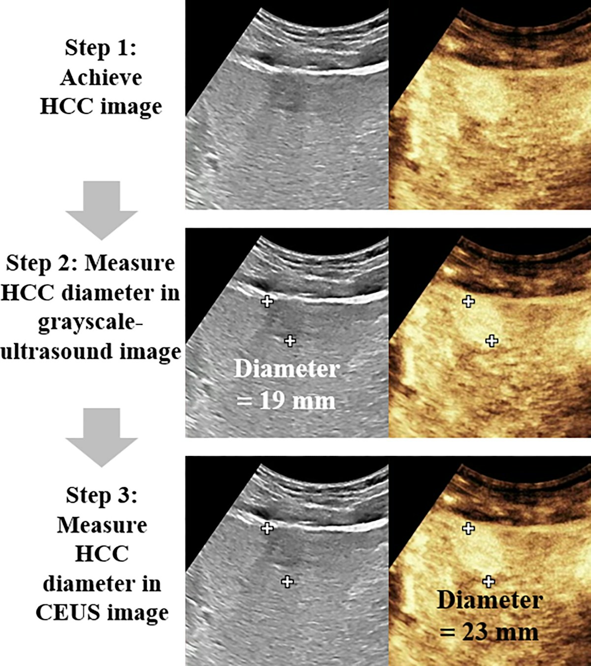
What the Beijing team measured
The study, led by Wenzhen Ding and Ran Ji of the Department of Interventional Ultrasound at the Chinese PLA General Hospital, starts from something every liver sonographer has seen: the outline of a nodule on gray-scale imaging does not always match the area that enhances after microbubble injection. Earlier studies had described this mismatch between vascularity and gross morphology, but, according to the authors, no one had defined it operationally or measured its clinical impact.
CEUS has the advantage of displaying both pieces of information in the same frame, side by side, with no image registration. The group defined PBM as a CEUS arterial-phase diameter exceeding the B-mode diameter by 5 mm. Measurement follows three steps: acquire the tumor plane, measure the largest diameter on gray-scale, then fine-tune the calipers on the contrast image. In the published example, a nodule in a 62-year-old man with cirrhosis went from 19 mm to 23 mm.
According to Diagnostic Imaging, the design was retrospective and multicenter, with a mean patient age of 60. The primary outcomes were recurrence-free survival (RFS) after laparoscopic hepatectomy and local tumor progression-free survival (LTPFS) after thermal ablation.
The numbers: recurrence, microvascular invasion and cytokeratins
Compared with patients without the sign, those with PBM had worse RFS after surgery (hazard ratio, HR, 1.76; p=0.03) and worse LTPFS after ablation (HR 2.40; p<0.001). The difference showed up in the surgical specimen too: microvascular invasion (MVI) was present in 52.2% of PBM tumors versus 25.0% of the rest (p=0.02). Cytokeratin-7 (CK7) positivity was 26.2% versus 11.3% (p=0.04), and cytokeratin-19 (CK19) positivity was 26.9% versus 8.8% (p=0.02).
The sign was more common in three groups: tumors larger than 3 cm (30.8%), recurrent HCC (28.4%) and cirrhotic livers (26.8%). The authors offer two explanations for the poorer prognosis. One is biological: MVI, CK7 and CK19 are established markers of aggressive behavior. The other is technical: if the operator plans treatment from the B-mode border, the true tumor extent is underestimated. That would explain why PBM had a larger effect on ablation, which depends directly on the image-defined margin, than on resection.
Technical context: why the margin matters in ablation
As background (not detailed in the news reports), radiofrequency or microwave ablation is a curative option for small HCC, and the working rule is to aim for an ablative margin of at least 5 mm of parenchyma around the tumor. Local progression typically arises at the periphery of the treated zone, where viable tumor foci escape the ablation. A nodule that “looks” 19 mm but has tumor perfusion out to 23 mm uses up nearly all of the planned margin before the needle goes in.
MVI, in turn, is one of the strongest predictors of early recurrence after resection or transplantation, but it can only be confirmed on histology. That is why there is so much interest in preoperative imaging predictors, such as the MRI score that predicts early liver cancer recurrence. PBM has a practical edge: it comes from an ultrasound exam, at the bedside or in the ablation suite, and needs no segmentation, postprocessing or dedicated software. Ultrasound platforms with elastography, CEUS and AI already provide the side-by-side dual mode the measurement requires.
Editorial: reproducibility is the selling point
In an accompanying editorial, Omid Shafaat and Stephen Reis of Columbia University in New York point out that five readers with no prior CEUS experience reached 92% accuracy in classifying PBM after just two hours of training, and that the finding held steady across different acquisition angles and time points. In their view, many imaging biomarkers never leave the lab because they depend on tedious segmentation, registration or complex postprocessing; a simple caliper-based criterion has a real chance of reaching routine practice. They conclude that, if prospectively validated, PBM could help bridge tumor characterization and image-guided therapy.
Practical implications
The authors suggest planning ablation margins from the CEUS-defined extent when PBM is present, and they argue for CEUS guidance particularly in patients with cirrhosis and recurrent HCC. The rationale: cirrhosis degrades border definition on B-mode but has little effect on CEUS, and many recurrent tumors are not candidates for repeat hepatectomy, which makes ablation the main curative option.
For programs in regions with large cirrhotic populations under ultrasound surveillance, including much of Latin America, where viral hepatitis, alcohol and fatty liver disease all contribute, the finding is relevant to hepatology and interventional radiology teams. Contrast ultrasound is still concentrated in referral centers in many countries, but adding one more measurement to a pre-ablation protocol costs little where CEUS is already in use. The sign may also weigh on the choice between ablation, resection and locoregional therapies such as transarterial embolization, an area where new devices like TriNav Advance for liver embolization aim to improve treatment delivery.
Limitations and next steps
The group acknowledges that the 5 mm threshold was derived and evaluated in the same cohort, which tends to overstate the marker’s performance. There was no direct comparison between ultrasound measurements and true size on the pathology specimen, and the criteria may need adjusting for asymmetric PBM or tumors abutting the liver capsule. The design is retrospective and comes from high-volume Chinese centers where hepatitis B predominates, which limits direct extrapolation to Western and Latin American cohorts. Prospective validation, ideally multicenter and with ablation planned from the CEUS margin, is the missing step.
Source: AuntMinnie, Diagnostic Imaging, Ding, Ji et al., Radiology 2026 and editorial by Shafaat and Reis



