

Adding pseudocapsule assessment to the clear cell likelihood score (ccLS) improved the separation of benign and malignant renal masses on multiparametric MRI. In a retrospective study from Gazi University in Ankara, Turkey, published in the European Journal of Radiology, the modified ccLS (m-ccLS) reached an area under the curve (AUC) of 77% for that task, compared with 65.4% for the standard ccLS, while keeping its accuracy for identifying clear cell carcinoma.
What the study evaluated
The team led by radiologist Umut Asfuroğlu of the Department of Radiology at Gazi University School of Medicine reviewed multiparametric MRI of 106 patients with renal masses, with a median age of 60.5 years. Most lesions (73 of 106) were cT1a tumors, meaning 4 cm or smaller, which is the group where choosing between surgery, ablation, biopsy or active surveillance is often hardest. The paper (Asfuroğlu U, Cindil E, Kalafat B, Badem A, Uçar M, Tokgöz N) appears in the October 2026 issue of the journal, DOI 10.1016/j.ejrad.2026.113288, and was covered by Diagnostic Imaging on October 7.
The researchers compared two versions of the score. The first is the standard ccLS, a five-point algorithm that estimates how likely a solid mass is to be clear cell renal cell carcinoma (ccRCC). The second is the m-ccLS, which adds the presence of a pseudocapsule as a criterion. They also analyzed quantitative parameters, including the T2 signal intensity (SI) ratio between the lesion and the renal parenchyma.
Key results
- Benign versus malignant: AUC of 77% for the m-ccLS and 65.4% for the ccLS, a gain of more than 11 percentage points.
- ccRCC versus non-ccRCC: similar performance for both versions (AUC of 88.4% for the ccLS and 86.8% for the m-ccLS). The modification did not give up the score’s original strength.
- T2 SI ratio: the most consistent single marker. It was higher in malignant than in benign lesions (1.10 vs. 0.85) and higher in ccRCC than in other types (1.16 vs. 0.85), with statistically significant differences in both comparisons.
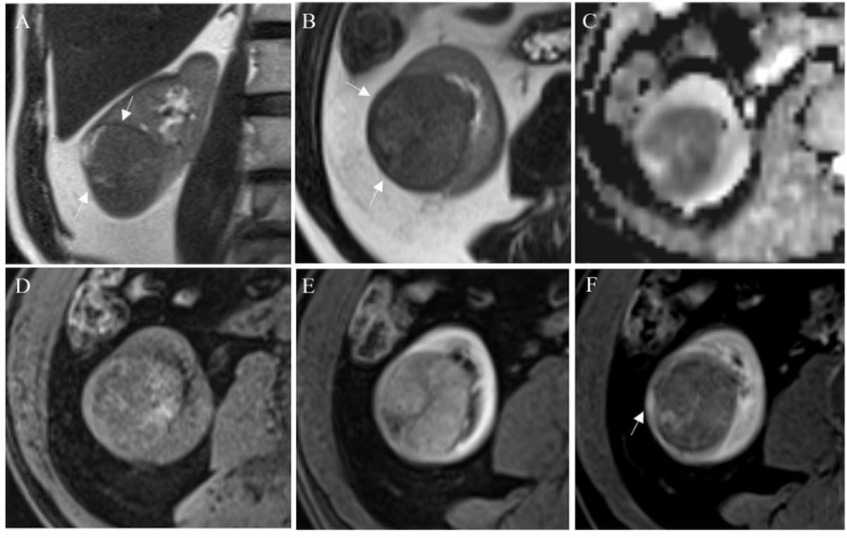
According to the authors, the explanation lies in non-clear cell subtypes. Papillary and chromophobe carcinomas tend to receive low ccLS scores, which can make them look like low-risk lesions. When the pseudocapsule is taken into account, some of these tumors are reclassified upward. The study’s own figure shows this: a chromophobe carcinoma that would have been ccLS 1 became ccLS 3 because of a well-defined capsule. The group notes that the T2 finding is consistent with the algorithm, since T2 signal is one of its core features.
Technical context: what the ccLS is and why the pseudocapsule matters
The ccLS was developed to bring structure to MRI reading of small solid renal masses, much as PI-RADS did for the prostate and LI-RADS for the liver. The logic is stepwise: the radiologist assesses T2 signal, the degree of corticomedullary-phase enhancement, microscopic fat on chemical shift imaging, segmental enhancement inversion and restricted diffusion, and arrives at a score from 1 to 5. High scores point to a high likelihood of ccRCC; low scores suggest other diagnoses such as papillary carcinoma or fat-poor angiomyolipoma. In the same vein of structured scores turning into decision tools, we recently covered an MRI score that predicts early liver cancer recurrence.
The known weakness is that the ccLS was designed to answer “is it clear cell?”, not “is it cancer?”. A papillary tumor, hypointense on T2 and only mildly enhancing, can land in the same low band as a benign lesion. The pseudocapsule, a fibrous rim of compressed parenchyma and reactive tissue usually seen as a hypointense ring on T2, is a sign of expansile growth frequently associated with renal cell carcinoma. Adding it may therefore help exactly where the original score is weakest. This line of research is not isolated: the Journal of Magnetic Resonance Imaging ran an editorial in February 2026 on another study that also integrates the pseudocapsule into the ccLS for small renal masses.
Practical implications
The literature commonly reports that a meaningful share of resected small renal masses turn out to be benign, exposing patients to unnecessary nephrectomies. A score that better flags benignity could help decide who can go to active surveillance or biopsy before surgery and who should move straight to treatment, including less invasive options such as percutaneous ablation, which performs comparably to surgery for small tumors.
The issue grows with incidental findings. The spread of whole-body MRI, such as the research network launched by Prenuvo, is likely to increase the number of renal lesions found in people without symptoms. On the molecular imaging side, CAIX-targeted girentuximab PET pursues another route to non-invasive ccRCC characterization. MRI with structured scoring, however, remains the most widely available tool.
In Brazil and across Latin America, where access to PET with new radiopharmaceuticals is limited, improving how an existing exam is read has immediate value. Pseudocapsule assessment needs no extra sequence: it can be done on the T2 and post-contrast images already in a standard renal protocol. That makes the change relatively easy to test in practices that already report the ccLS.
Limitations and next steps
The authors list the constraints themselves. This is a retrospective single-center study with a relatively small cohort and few benign lesions, which reduces the precision of the AUC estimates for the benign-versus-malignant task, precisely where the m-ccLS stood out. In addition, not every lesion had histopathologic confirmation. The coverage also does not make clear how much inter-reader agreement changes when a subjective criterion such as the pseudocapsule is added.
Before any change to routine practice, the m-ccLS needs multicenter, prospective validation with readers of varying experience and a head-to-head comparison against biopsy. Even so, the study reinforces two useful messages for anyone reading renal MRI today: the pseudocapsule deserves systematic description, and the T2 SI ratio may be a simple, reproducible quantitative complement to the score.
Source: Diagnostic Imaging



