

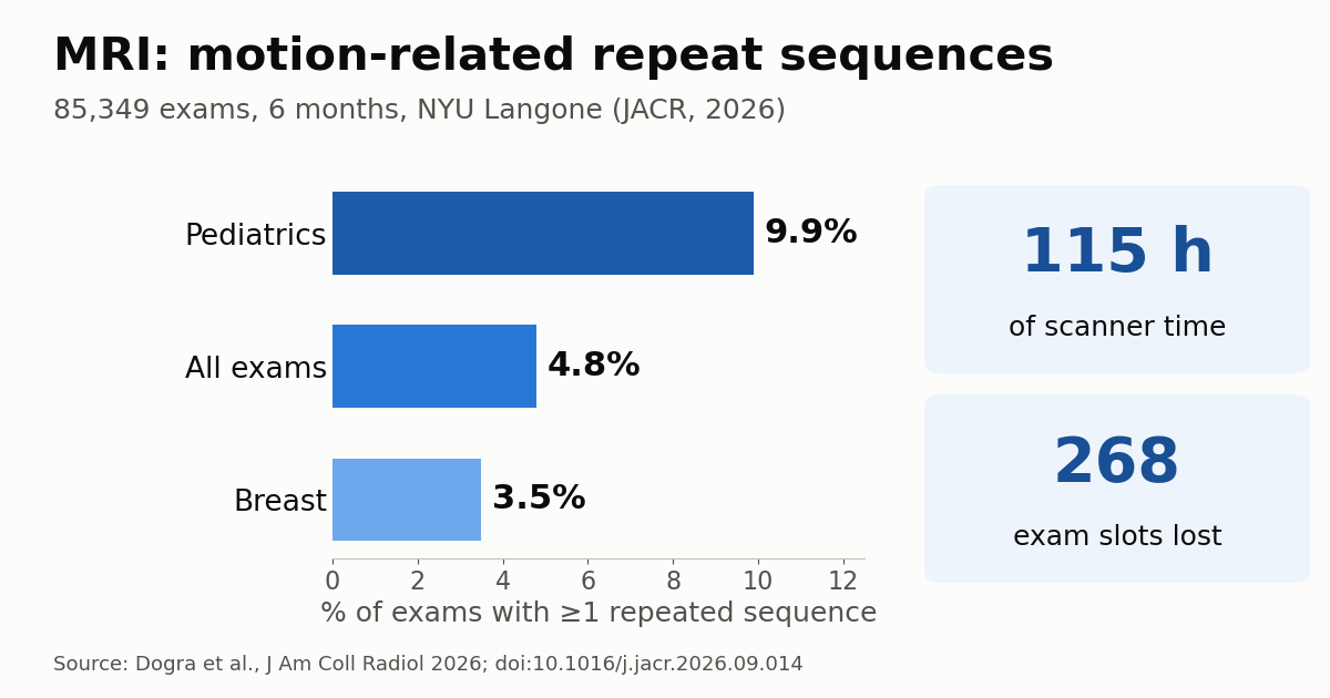
Repeating MRI sequences because of patient motion consumed 115 hours of scanner time over six months, the equivalent of 268 lost exam slots, across a large New York health system. The study, published in the Journal of the American College of Radiology (JACR) by radiologists at NYU Grossman School of Medicine working with Siemens Healthineers, analysed 85,349 MRI exams and found at least one repeated sequence in 4.8% of them. The more useful finding is a different one: repeating does not always help. In brain MRI a repeat almost always improved the image; in MR cholangiopancreatography (MRCP) it produced no statistically significant gain.
What the NYU study measured
The paper, led by Siddhant Dogra with Hersh Chandarana as senior author, is retrospective and covers six months of exams on scanners with sequence-level analytics, meaning every acquisition within an exam is logged. The team identified exams in which the technologist had flagged a sequence as a repeat. Three co-authors are from the Magnetic Resonance division of Siemens Healthineers in Erlangen, Germany.
Of the 85,349 exams, 4,072 had at least one sequence re-acquired. Frequency varied widely by segment: highest in pediatrics (9.9%) and lowest in breast MRI (3.5%). According to The Imaging Wire’s write-up, repeat time added up to 115 hours of scanner time, or 268 schedule slots. Running the numbers backwards ourselves gives roughly 1.7 extra minutes, on average, per affected exam. That sounds small until it is multiplied across a high-volume enterprise.
The second part of the study was a reader evaluation. For brain MRI, four readers compared 87 baseline-repeat pairs from 77 patients, scoring image quality, diagnostic confidence and whether they would request yet another repeat. Repeats improved every metric significantly (p<0.001), but 19.5% of repeated sequences did not improve, and the gain shrank as elapsed exam time grew. For MRCP, two readers assessed 30 pairs from 27 patients: repeats did not significantly improve quality or confidence, and both readers would have asked for another repeat in half of the cases.
Why the brain responds and MRCP does not
The difference makes physical sense. In the brain, motion is usually voluntary and episodic: the patient turns the head, swallows, shifts position. A fresh acquisition, with better coaching and immobilisation, has a good chance of coming out clean. Brain sequences are also relatively short, and robust motion-tolerant options exist, such as radial k-space sampling (PROPELLER/BLADE).
MRCP relies on heavily T2-weighted 3D sequences, usually respiratory-triggered. The artefact comes from irregular breathing, peristalsis and trouble holding a breath, factors that recur on the next attempt. When the cause is physiological and persistent, re-running the same sequence tends to reproduce the same problem, only with more table time. The authors do not break down the mechanism, but the result supports a clear reading: the decision to repeat should depend on anatomic region and sequence type, not on reflex.
The falling benefit with exam duration also has an intuitive explanation, which is ours rather than the authors’: a tired, uncomfortable or anxious patient many minutes into the bore is unlikely to hold stiller on the third try.
Is 4.8% low? The literature context
The rate is lower than earlier studies of motion-related repeats, which ranged from 7.5% to 30%. A widely cited University of Washington paper, also in JACR, in 2015 estimated repeat sequences in about 20% of exams and forgone revenue on the order of $115,000 per scanner per year.
Two explanations are plausible, and both appear in The Imaging Wire’s analysis. The first is methodological: the study counts only technologist-labelled repeats, which likely underestimates the total because not every repeat gets flagged. The second is technological: over the past decade departments have adopted shorter protocols, parallel imaging, compressed sensing and deep learning reconstruction, all of which cut acquisition time and, with it, the window for motion.
What this changes for MRI operations
The authors’ main recommendation is to track repeats at the sequence level, not just the exam level. With that data a manager can see which protocol, scanner and shift loses the most time, and respond in a targeted way: better patient preparation, swapping a Cartesian sequence for a radial one, shortening a protocol, or setting rules for when a repeat is not worth it.
For busy outpatient and hospital sites alike, 268 slots in six months represent patients who could have been scanned. We have reported that MRI efficiency gains could lift revenue per scanner by up to 60% and that AI tools can cut MRI wait times by more than 50%. Measuring repeats is a cheap step that should come before either initiative. In Brazil and much of Latin America, where scanner hours are expensive and public-system waiting lists are long, the argument is even stronger.
Pediatrics deserves special attention. At nearly 10% repeats, it has the most to gain from fast protocols, immobilisation and child-friendly preparation, as discussed in our coverage of rapid MRI for pediatric traumatic brain injury. The human factor also matters: the technologist shortage covered in our piece on DeVry’s new MRI degree directly affects the quality of patient coaching, the first line of defence against motion.
Limitations and next steps
This is a single-system study, on one vendor’s scanners, with that vendor among the authors, which limits generalisability. Reliance on manual technologist labelling is the most important limitation because it understates the true rate. The reader-study samples are also small, particularly for MRCP at 30 pairs, so claims that repeats “don’t work” there should be made with caution.
Even so, the message is practical: motion still costs real scanner time, and not every repeat buys quality. Prospective motion correction, external tracking sensors and AI reconstruction should keep shrinking the problem. Until then, knowing where repeats happen, and where they are not worth it, is an operational win in itself.
Source: The Imaging Wire; original study: Dogra S et al., J Am Coll Radiol, 2026



