

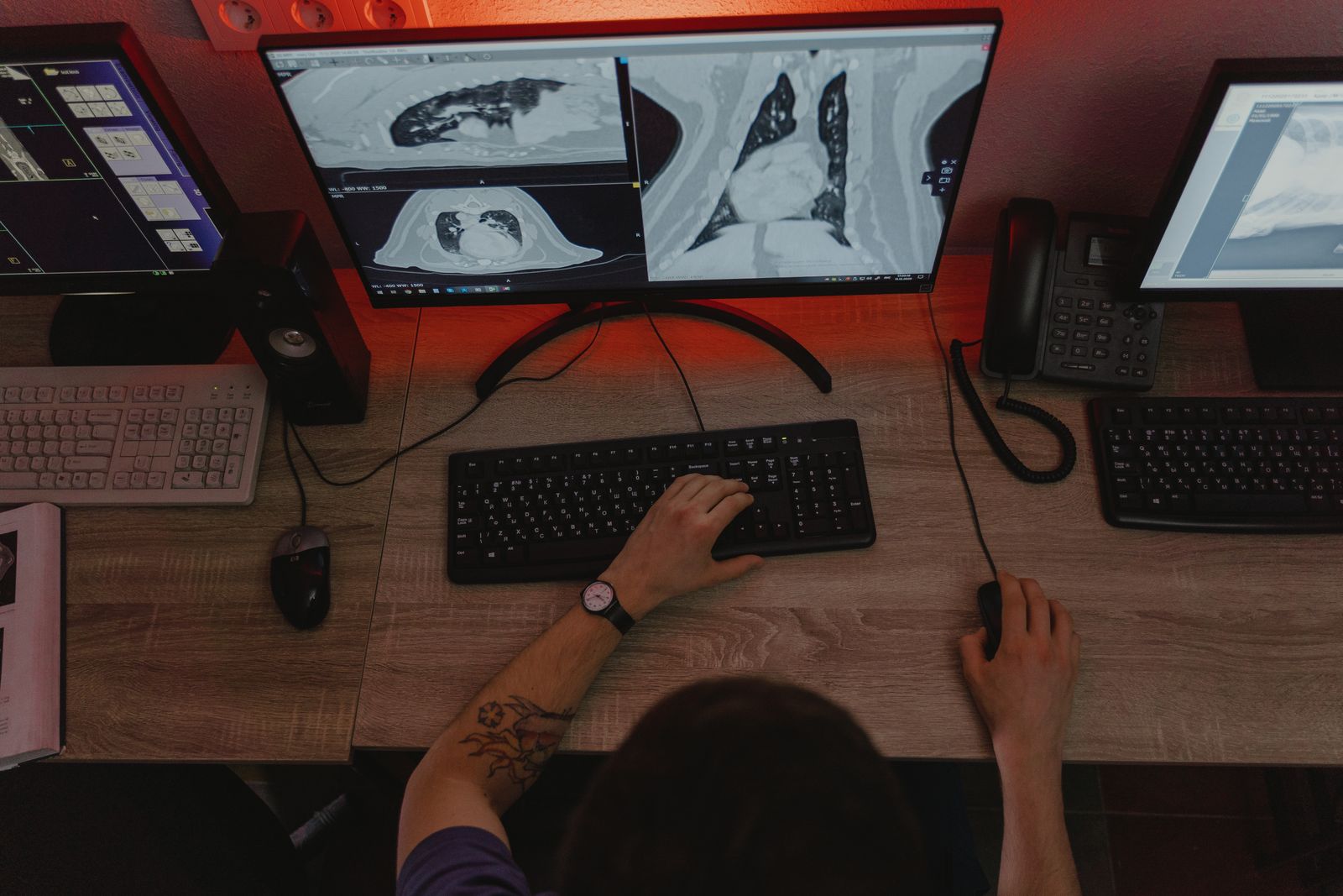
The display is now quality infrastructure, not a peripheral
Diagnostic monitors used to be judged on a short list of numbers: resolution, luminance, contrast, uniformity, grayscale behaviour. Those numbers still matter, but a feature in Radiology Today argues they no longer describe what departments are actually buying. Vendors have stopped selling panels and started selling reading environments — built-in front photometers, automated calibration software, fleet-wide quality management and ergonomics priced as one package.
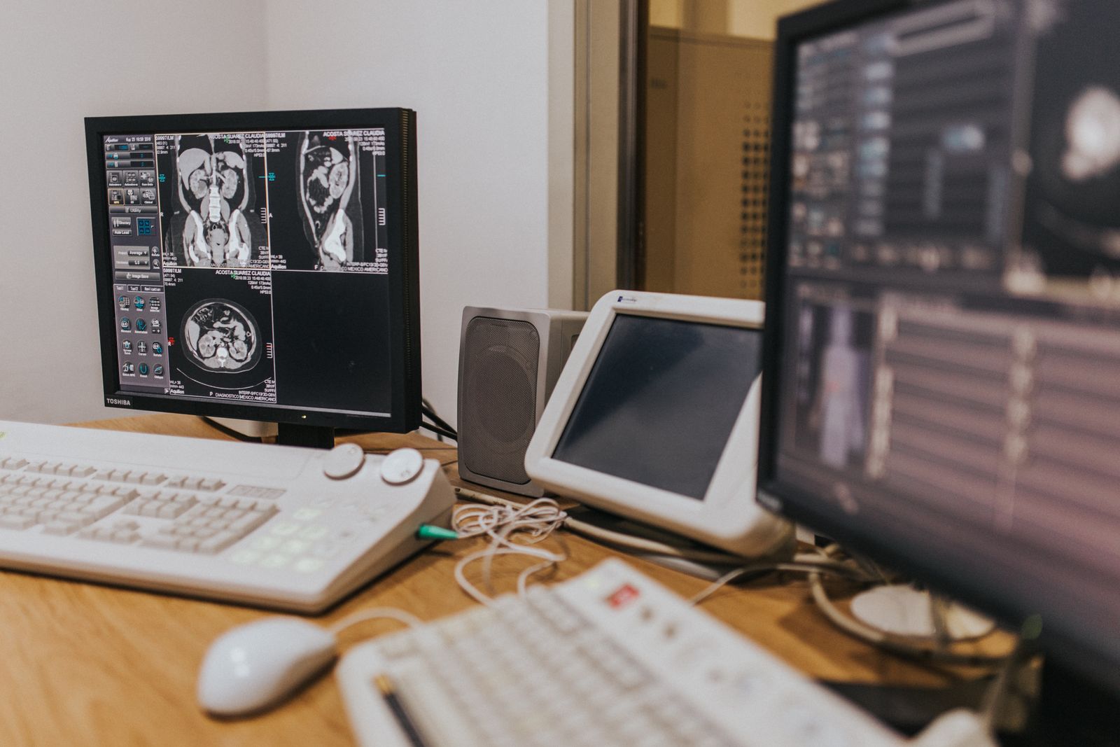
What forced the change is geography. A single radiologist may read from a hospital reading room, an outpatient imaging centre and a home office inside one week, and image quality cannot be a function of which desk they happen to sit at. The purchasing question shifted accordingly. It is no longer which monitor has the most pixels, but how a health system proves — on demand, in an audit — that every screen in the network is still inside specification.
What the vendors interviewed are seeing
Tara Neill, director of sales and marketing at Double Black Imaging, frames the shift around integration rather than hardware: front sensors, calibration software and enterprise management tools now operate as one system, and the display has stopped being treated as standalone equipment. Her operational point is blunter. A monitor has to perform on the day it is installed and every day after that, and one-off manual inspection does not scale across hospitals, clinics and radiologists’ homes.
Remote breast imaging is the pressure test. As telemammography expands, stable luminance, consistent grayscale rendering, accurate colour and documented quality assurance have to exist wherever the study is opened, not only in the main reading room. Double Black’s CFS Calibration Software is aimed squarely at that gap, automating DICOM calibration, conformance testing, reporting, alerting and fleet management so PACS, IT and compliance staff can see a drifting screen before it reaches a report.
Jim Salamon, senior health care IT manager at LG Electronics USA, adds the AI dimension. Measurements, annotations, segmentations, probability scores, colour overlays, heat maps and worklist priority flags now compete for the same screen real estate as the primary image. The display has become the point where a radiologist decides whether to act on an algorithm’s output, and clumsy presentation erodes trust in the tool long before anyone argues about its sensitivity.
On configurations the article records a genuine split rather than a winner. Large-format 6, 8 and 12 megapixel screens are gaining ground because they remove bezels from the visual field, while dual 3 and 5 megapixel setups stay popular because they can be angled inward and match established reading habits. KVM switching, mounts, sit-stand desks, task lighting and management software all landed on the same purchase order. JVC and Barco are pushing the large-format route with built-in calibration, luminance stabilisation and uniformity correction, Barco’s Coronis line combining grayscale and colour on one panel for multimodality work.
Why the DICOM GSDF is what makes two screens agree
Calibration here has a precise technical meaning. Part 14 of the DICOM standard defines the Grayscale Standard Display Function, a curve that maps every grey value sent by the viewer to a luminance measured in candelas per square metre. It is not an arbitrary gamma. It is derived from Barten’s model of human vision and spaces grey levels in just-noticeable differences, so that one step of pixel value produces roughly the same perceptual change anywhere on the scale, from the darkest to the brightest region of the image.
The function is defined across a luminance range of 0.05 to 4,000 cd/m², interpolated over 1,023 levels derived from that model. Calibrating a display to the GSDF means bending the panel’s native response onto that reference within a tolerance. That, and not matching hardware, is why two radiologists in different cities can discuss the same window setting and mean the same thing. It is also why a distributed reading operation is only as consistent as its worst-calibrated endpoint, a theme we explored in our piece on cloud teleradiology and distributed reading.
The numbers that turn this into a purchasing specification come from the ACR–AAPM Technical Standard for Diagnostic Interpretation Displays, revised in 2024, and from AAPM Report 270, the successor to the long-serving TG-18. The standard recommends a combined maximum luminance of at least 350 cd/m² for general interpretation and 420 cd/m² for mammography, with minimum luminance of 1.0 and 1.2 cd/m² respectively. The target luminance ratio — maximum over minimum — is 350, with an acceptable band of 250 to 450, a figure anchored in the contrast of a chest film on a lightbox.
Conformance is measured rather than assumed. Using 18 equally spaced points, every 15 grey levels on an 8-bit system, the standard allows a maximum error of ±10% from the GSDF. Finer 52-point sampling exposes calibration defects better but needs its own pass criterion. Ambient illuminance should sit between 25 and 75 lux, and reflected ambient luminance below a quarter of the display’s minimum luminance — a clause that alone disqualifies most improvised home setups. Uniformity is checked at acceptance, with deviation under 15%.
The same standard tempers enthusiasm for built-in sensors. They sit at the panel edge and read a different luminance from the centre, so they must be correlated with an external photometer at installation and, per Report 270, every 10,000 backlight hours. Relying solely on vendor software to verify performance is explicitly discouraged. Layered on top are visual checks — TG270-sQC or TG18-QC patterns reviewed at least quarterly by trained staff — and annual quantitative testing under the general supervision of a qualified medical physicist.
What changes at the workstation
For a department, the practical consequence is that display QA stops being a facilities task and becomes an imaging informatics programme with an owner, a schedule and an audit trail. Acceptance testing when a station goes live. A declared calibration target per clinical use. A quarterly pattern review that someone actually performs and signs. Luminance history and exception reports that survive an accreditation visit. None of this is new science, but doing it across a hundred endpoints without centralised software is arithmetic that does not work.
The home reading station is where most programmes break. A consumer monitor bought by the radiologist, running a factory curve in a room with uncontrolled light, will not reproduce the dark end of the image the way the hospital screen does, and nobody will notice until a subtle finding is missed. Fixing it is not glamorous: enrol the endpoint in the same management console, set the same calibration target, verify it remotely, and replace the hardware on a planned cycle rather than after it fails.
Ergonomics belongs in the same conversation for a reason that is economic rather than sentimental. Stable luminance, antiglare treatment, ambient light awareness and sensible mounting reduce the number of times a reader manually adjusts an image or stops to wonder whether a perceived difference is clinical or a display artefact. Over a heavy shift that friction accumulates, which is precisely the mechanism described in our coverage of musculoskeletal strain among radiologists.
Software beats panels, at least for now
The article is deliberately unexcited about panel technology. OLED, mini-LED and micro-LED may eventually reshape diagnostic displays, but they first have to demonstrate stability, calibration capability and long-term consistency, because in health care a newer panel only matters if it still meets specification years later. The expectation set out by both vendors is that the next wave of progress arrives in management software rather than in physics.
Digital pathology will widen the argument further, since whole-slide reading depends on colour fidelity and stain differentiation rather than grayscale alone, and colour QA has far less institutional muscle memory behind it than luminance QA. Add AI overlays that use colour to mark suspected findings, and chromaticity stops being a cosmetic specification.
The budget lesson is the one worth carrying into a tender. The cost of a reading environment is not the price of the screen: it is graphics controllers, mounts, photometers, calibration software, furniture, installation and ongoing service, plus a predictable replacement cycle. Replacing monitors reactively after failure is the most expensive model available, because the interval between silent drift and visible failure is exactly the period during which studies were reported off specification.
Source: Radiology Today



