

The First Achromatic Neutron Lens Already Produces Magnified Images
A team at the Paul Scherrer Institute (PSI) in Switzerland has built the first achromatic lens for neutrons and used it to capture magnified images with resolution below 20 micrometers — including from objects sitting six meters away from the detector. The work, led by Mano raj Dhanalakshmi Veeraraj and published in Nature Communications, clears the obstacle that had blocked neutron microscopy since the first attempts to focus such beams: chromatic aberration.
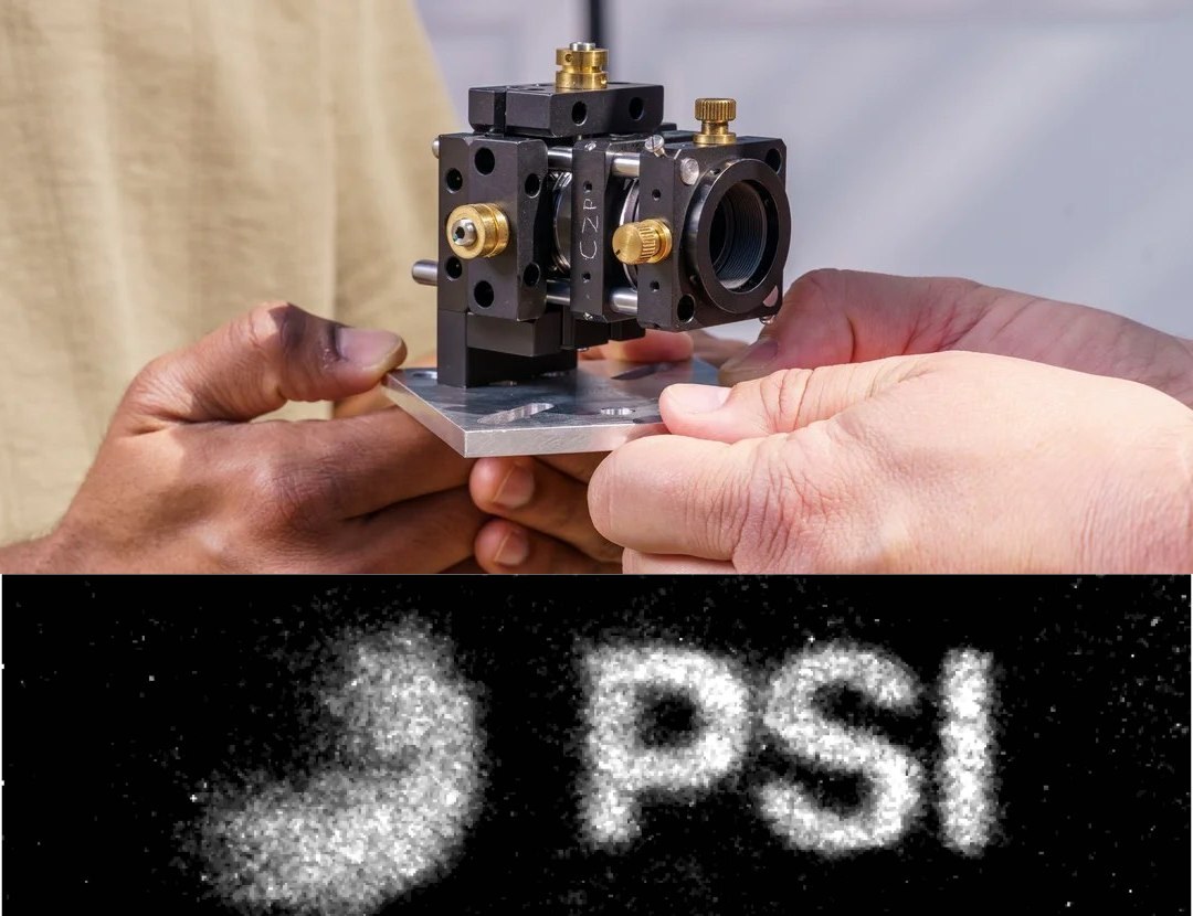
It is worth spelling out why this matters to anyone working with imaging. Neutrons and X-rays are complementary, not competing. X-rays interact with the electron cloud and therefore see dense, heavy material well; neutrons interact with the nucleus and are exquisitely sensitive to light elements — hydrogen and lithium above all. The practical consequence is elegant: a neutron crosses tens of millimeters of metal and still resolves the oil inside the engine, the polymer inside the housing, the lithium inside the battery. The same contrast mechanism lets researchers follow water uptake in plants and examine archaeological artifacts without destroying them.
Why Focusing Neutrons Is So Hard
The difficulty arises from the very property that makes neutrons useful. Electrically neutral and weakly interacting, a neutron penetrates deep — and resists being deflected. Its refractive index departs only marginally from unity:
$$n = 1 – \frac{\lambda^{2} N b_{c}}{2\pi}$$
where $\lambda$ is the neutron wavelength, $N$ the number density of nuclei and $b_{c}$ the coherent scattering length. For thermal and cold neutrons, the deviation $1-n$ sits in the range of $10^{-5}$ to $10^{-6}$ — refraction that is essentially negligible next to visible optics. That is why a “neutron lens” could never be a curved block of glass.
The second obstacle is the beam itself. Neutron sources are polychromatic and of low brightness: the beam carries many wavelengths at once, and discarding nearly all of them with a monochromator means throwing away the little flux available. Yet the known optical elements disperse. Compound refractive lenses (CRLs) and Fresnel zone plates (FZPs) had already been demonstrated for neutron imaging, but with severe chromatic aberration under polychromatic beams — each wavelength focuses in a different plane, and the image is hopelessly blurred.
The Achromat Trick: Nickel Diffracts, Diamond Refracts
The PSI solution is the same idea that solved achromatization in visible optics three centuries ago, transposed into a completely different physical regime: combine two elements whose dispersions cancel out. The lens consists of concentric nickel rings — the Fresnel zone plate, which works by diffraction — added to structures precisely sculpted in diamond, which work by refraction, all arranged in a carefully defined geometry.
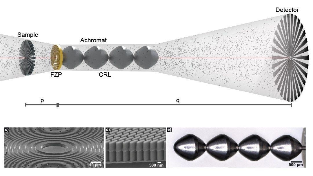
Why it works comes down to how each element depends on wavelength. In a zone plate, focal length scales as $f_{FZP} \propto 1/\lambda$. In a compound refractive lens for neutrons, since $1-n \propto \lambda^{2}$, focal length scales as $f_{CRL} \propto 1/\lambda^{2}$. The two dispersions carry different signatures — and by choosing the right proportion between the elements, the first-order dependence on $\lambda$ cancels. What remains is a system that focuses a broad band of wavelengths at the same point, exactly what a low-brightness polychromatic beam demands.
Material choice is not decorative. Nickel provides suitable phase contrast for the diffractive zone in neutrons, while diamond combines low absorption with the hardness and stability needed to hold microstructures machined to sub-micrometer precision. Fabricating that is high-aspect-ratio lithography at the edge of the state of the art — and much of the paper’s technical merit lives there.
Why Detector Distance Changes Everything
Here is the most underrated gain in the paper. Conventional neutron radiography works in pinhole geometry: the beam is collimated by an aperture of diameter $D$ at distance $l$ from the sample, and geometric penumbra in the image grows linearly with the sample-to-detector distance $L$,
$$u_{g} = \frac{D \, L}{l}$$
which imposes a cruel rule: to preserve resolution, the sample must sit almost against the detector. That rules out any large object and any sample inside a cryostat, pressure cell or environmental chamber — precisely the most interesting experiments in materials science and engineering.
The lens breaks that constraint. In the first tests, the researchers imaged a commercial lithium-ion battery positioned six meters from the detector and magnified the layered electrode assembly sevenfold. Because the image arrives magnified, spatial resolution is no longer dictated by detector pixel size — the same reasoning any medical physicist recognizes when discussing pixel pitch and modulation transfer function in digital radiography. “If you have a long enough beamline, in principle you could magnify more. The magnification isn’t limited by the lens, but by the length of the instrument,” Veeraraj said.
What This Has to Do With Medical Physics
A clinical reader may reasonably ask what a neutron lens has to do with daily radiology. At least three things. The first is conceptual: this is another case where image quality improved through optics and geometry rather than by raising flux — the same logic behind the debate over when a noisy image is already good enough for diagnosis in CT. Resolution obtained by design costs less than resolution obtained by dose.
The second is instrumental. Neutron beams are the reference tool for characterizing materials that radiology and radiotherapy use without a second thought: hydrogen in plastics and anthropomorphic phantom polymers, lithium in detectors, water uptake in tissue substitutes, integrity of accelerator components. Neutron microscopy with genuine magnification widens the repertoire of anyone doing materials quality control.
The third is methodological. Neutron transport is the same computational physics problem that underpins Monte Carlo dose calculation — the method was born precisely to solve neutron diffusion before migrating to dosimetry. Anyone who has compared pencil beam and Monte Carlo algorithms for proton dose calculation knows the terrain. And as proof that particle imaging keeps reinventing itself in hostile environments, recall the first diagnostic radiograph acquired in space on the Fram2 mission.
Limits and Next Steps
Expectations need calibrating. Neutrons require a reactor or a spallation source — there is no benchtop version, and the queue for beamtime at facilities such as PSI is long. Twenty-micrometer resolution is excellent for neutrons but remains far behind what synchrotron X-ray microscopy delivers. Achromat efficiency across the full spectral band, usable field of view and tolerance to misalignment are exactly the parameters the next generation of the device will have to improve.
The authors are explicit about how early this result is. “This is just the beginning. We already see ways to improve the lens. The crucial point isn’t simply resolution, but a completely new way of capturing images,” Veeraraj said. That is the right reading: the paper does not deliver a finished technique, it delivers a class of instrument that did not exist before — and opens a path toward high-resolution neutron microscopy for experiments that until now had to choose between magnification and sharpness.
Source: Inovação Tecnológica | Original paper: An achromatic neutron lens, Nature Communications



