

One Third: The Share of U.S. Prostate Workups That Include MRI
Only about one third of prostate cancer workups in the United States include an MRI before biopsy — while Australia, Canada, the U.K. and much of Europe have already adopted pre-biopsy MRI as the standard of care. A recent STAT report put the gap back on the table and revived an uncomfortable question: if the evidence supports it and the medical societies recommend it, what is holding adoption back? The answer has three layers — training, access and money.
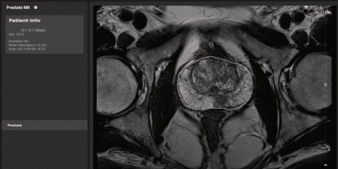
The underlying clinical problem is an old one. PSA alone is a poor trigger: it flags who deserves attention but does not separate the cancer that will kill from the finding that would never have mattered. Traditional systematic transrectal biopsy, which typically takes 10 to 12 cores across a fixed template, inherits that ambiguity and adds two problems of its own — it samples the gland blind, and it charges a price in discomfort, bleeding and infection risk.
93% Versus Roughly 50%: The Number Behind the Shift
This is where MRI enters. Compared with systematic biopsy, multiparametric MRI is noninvasive and substantially more sensitive for clinically significant lesions: about 93% versus something near 50%. Those figures trace back to the PROMIS trial in The Lancet, which compared multiparametric MRI and transrectal ultrasound-guided biopsy against template mapping biopsy as reference. That result is what opened the door to protocol change in Europe: MRI misses the important tumor less often.
The second pillar came from PRECISION, in the New England Journal of Medicine, which tested the full strategy — MRI first, biopsy targeted only at suspicious lesions — against standard systematic sampling. The image-guided pathway detected more clinically significant cancers while finding fewer low-grade ones, the kind that generate surveillance, anxiety and repeat procedures without changing outcomes. Reporting is standardized through PI-RADS, which scores each lesion from 1 to 5 by combining T2-weighted imaging, diffusion with its ADC map and, when indicated, dynamic contrast-enhanced sequences.
Training, Access and an Inverted Financial Incentive
The STAT report groups the U.S. obstacles into three fronts. The first is education: many urologists were never trained to interpret a prostate MRI report or to perform fusion-guided targeted biopsy. Without that skill, the scan becomes information nobody knows how to use — and an exam that does not change management is, in practice, a disposable exam.
The second is access. High-quality MRI capacity, with a dedicated protocol and a radiologist experienced in prostate reads, is not evenly distributed. The disparities are explicit: Black patients, people living outside cities and Medicaid-eligible populations are less likely to reach the scan. Scheduling matters as much as geography here — when the MRI calendar is saturated, elective prostate imaging is the first to slip, and optimization tools have already cut MRI wait times by more than half in departments that attacked the scheduling problem directly.
The third front is the least comfortable. Biopsies are often performed in-house at the urology practice, with reimbursement that can reach $1.5k per procedure. Systematic biopsies also detect low-grade cancers that might never become clinically significant — cancers that feed active surveillance and repeat biopsies, generating further revenue. Wider MRI use would shrink that stream. This is not an accusation of bad faith; it is an incentive structure pointing away from the evidence, and it operates in the same economic climate that made the 2027 Medicare fee schedule such a flashpoint for imaging revenue.
What the Societies Say — and What Payers Cover
On the recommendation side, the picture is friendlier than practice suggests. The National Comprehensive Cancer Network endorses pre-biopsy MRI; the American Urological Association and the American Society of Clinical Oncology do as well, if conditionally. A 2020 paper was decisive in persuading insurers to cover prostate MRI — and a share of clinicians simply do not know the coverage exists. That is a quiet waste: the barrier many cite as financial has already been partly dismantled on paper.
MRI is not confined to the decision to biopsy, either. It informs staging, the selection of candidates for focal therapy and surgical planning. We have covered how MRI can take over part of the digital rectal exam’s role in local staging. Each additional use raises the marginal value of the same scan — a relevant argument for anyone who has to justify buying magnet time.
What Would Move the Needle
The proposed levers are pragmatic. Training embedded in urology residency, to close the competency gap at the source. Education for urologists and patients about reimbursement options and clinical benefit. Better access to high-quality MRI outside major metropolitan centers. And above all, unified implementation-focused programs: the Czech Republic’s trial, which combined a single protocol with training and communication, raised pre-biopsy MRI utilization by 10 percentage points. Recommendations alone do not change behavior; structured programs do.
Some limits deserve acknowledgment before treating MRI as a complete solution. Exam quality depends heavily on hardware, protocol and reader experience, with well-documented interobserver variability for PI-RADS 3 lesions. A negative MRI reduces but does not eliminate the chance of clinically significant cancer, and the biparametric approach — dropping gadolinium to save time and cost — still draws debate about sensitivity in specific scenarios. None of this invalidates the workflow change; it simply means the change demands investment in standardization, not just in equipment.
For radiology departments, the strategic reading is straightforward. Prostate MRI is a growing service line whose bottleneck is rarely the magnet itself: it is protocol discipline, structured PI-RADS reporting and a working relationship with urology. Departments that solve those three problems will capture the demand when the guidelines finally translate into routine — and the U.S. gap versus Europe suggests that translation is a matter of when, not whether.
Source: The Imaging Wire



