

Artificial intelligence may soon remove the need for gadolinium contrast in many cardiac MRI scans. A multicenter study published in the Journal of the American College of Cardiology (JACC) found that an AI tool called Virtual Native Enhancement (VNE) can reconstruct images equivalent to late gadolinium enhancement from sequences already acquired without any injection, spotting myocardial scars with roughly 94% accuracy when the images are high quality.
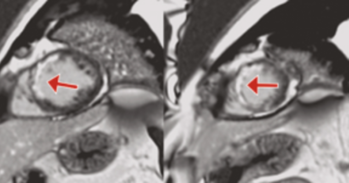
For radiologists and cardiologists, the finding raises the prospect of reserving contrast agents only for the patients who truly need them, cutting cost, scan time and the burden of an intravenous injection without losing diagnostic information. That same drive toward leaner imaging workflows is already reshaping practice, where AI has cut MRI wait times by more than 50% in the services that adopted it.
What the VNE study showed
This was the first prospective, multicenter and blinded validation of the technology in real clinical settings. Researchers enrolled 136 patients with a history of heart attack across three referral hospitals: the University of Oxford and the University of Leeds in the United Kingdom, and Fuwai Hospital of the Chinese Academy of Medical Sciences in China. Using only cine images and native T1 maps acquired before any contrast, VNE generated virtual images that mimic late gadolinium enhancement (LGE).
The synthetic images were scored by readers blinded to clinical data and to the actual contrast-enhanced scan. When VNE produced images rated high quality and high confidence, agreement with the reference standard was strong: the tool identified myocardial scars with about 94% accuracy. Readers judged that nearly 70% of the cohort had high-quality, high-confidence images, suggesting that a large share of patients could undergo cardiac MRI without gadolinium while preserving diagnostic quality.
The multicenter design was deliberate. “A major problem with medical AI is that systems which work well in one hospital often fail when tested somewhere else,” said senior author Stefan Piechnik, PhD. “We designed this study very carefully to make sure the scans were performed consistently and the AI was tested fairly in real clinical settings.” VNE was developed at Oxford’s cardiovascular magnetic resonance center (OCMR), and an earlier validation in hypertrophic cardiomyopathy had already been published in Circulation in 2021.
Why gadolinium matters in cardiac MRI
In current practice, late gadolinium enhancement is the reference test for detecting fibrosis and scarring in the heart muscle. After an intravenous injection, the contrast accumulates in damaged or fibrotic tissue, which lights up on images taken a few minutes later. This scar map guides important clinical decisions, from arrhythmia risk to therapy planning in patients who have already had a heart attack.
The trouble is that gadolinium carries costs and caveats. There is the rare but serious risk of nephrogenic systemic fibrosis in patients with kidney failure, along with evidence of metal retention in tissues such as the brain and bones after repeated exams. Add to that the cost of the agent, the need for venous access, the extra scan time, the risk of allergic reactions and the restrictions in pregnant patients and those with impaired renal function. Removing contrast when it is not essential makes the exam safer and simpler to run. Industry is chasing safer options too: the FDA recently cleared a low-dose MRI contrast agent from Bayer, a sign that reducing gadolinium exposure is a sector priority.
Who really needs contrast?
The most immediate value of VNE is not to replace gadolinium in every case, but to triage who genuinely needs it. If the AI delivers reliable images in nearly 70% of patients, a service can focus contrast injection on the minority in whom the virtual reconstruction falls short. “This technology could make heart MRI scans quicker, simpler and easier for many patients by reducing the need for contrast injections,” said senior author Vanessa Ferreira, MD, PhD. “Our long-term goal is to make cardiac MRI as straightforward and accessible as an ultrasound heart scan.”
The implications reach beyond a single scanner. Fewer injections mean fewer cannulations, shorter table times and less consumable spend, which in turn frees capacity for more patients. In health systems where advanced cardiac imaging is scarce or expensive, a protocol that skips gadolinium for a large fraction of exams could widen access, particularly where contrast supply and nursing staff for venous access are limited. For clinicians, it is a chance to offer myocardial scar assessment to more patients without overloading the service.
What still needs validating
The authors themselves are cautious. Larger studies are still needed before VNE can broadly replace contrast-enhanced cardiac MRI, and roughly a third of the study patients would still rely on the conventional exam to secure image quality. The critical issue is generalization: an AI that shines at one center can stumble at another, with different scanners, protocols and populations. That is precisely why the blinded, multicenter design mattered so much.
This caution echoes a wider debate about trusting algorithm-generated images. Synthetic imaging can be convincing enough to fool even experienced radiologists, which reinforces the need for rigorous validation and human oversight before any clinical decision. If upcoming trials confirm the results, VNE could become a valuable tool for deciding, scan by scan, which patients truly need gadolinium, moving cardiac MRI a step closer to the simplicity of an ultrasound.
Source: Radiology Business



