

MRI reveals higher neuro symptom burden in former soccer players
Former elite professional soccer players already show, in midlife, a higher burden of neuropsychiatric symptoms and structural brain changes visible on magnetic resonance imaging (MRI), according to research presented on July 12, 2026 at the Alzheimer’s Association International Conference (AAIC) in London. The study compared 142 former athletes with 56 healthy controls and found more depression, more anxiety and lower gray matter volume in regions tied to memory, attention and emotional regulation, even before any clinically apparent neurodegenerative disease.
How the study was conducted
The research was led by Caleigh Grace Lynch, of the Department of Brain Sciences, and Dr. Thomas D. Parker, a clinical lecturer in neurology, both at the UK Dementia Research Institute Centre for Care Research and Technology at Imperial College London. The work is part of the Advanced BRAIN Health Clinic research programme at the Institute of Sport, Exercise and Health and was funded by the Football Association (FA) and the Professional Footballers’ Association (PFA), the governing bodies of British professional football.
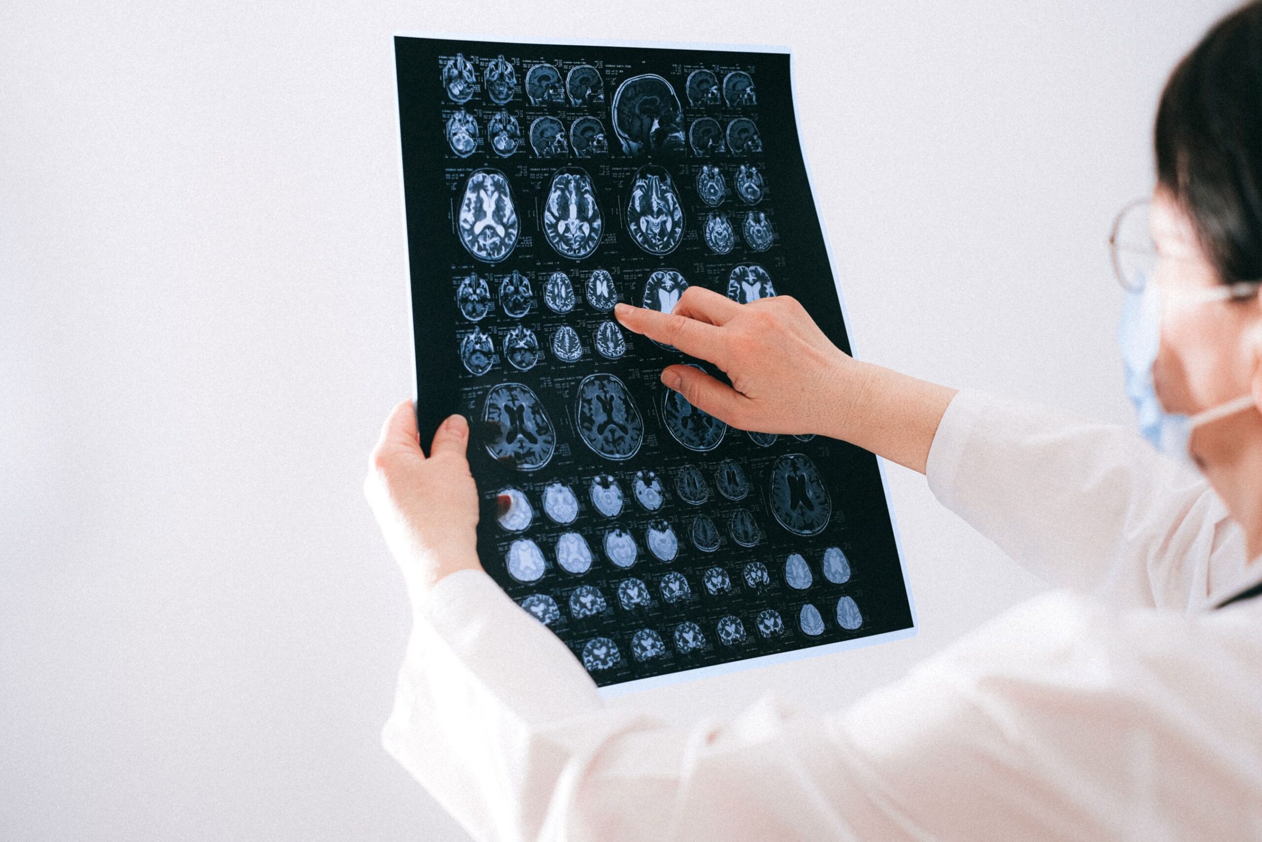
The team assessed 142 former professional players (126 men and 16 women), aged 30 to 60, compared with 56 control participants (43 men and 13 women) with no history of contact sports or repeated head impacts. Every participant completed standardized depression and anxiety assessments, objective cognitive testing and self-rated measures of functions such as planning, focus and problem solving. A total of 124 former athletes underwent brain MRI, with the scans reviewed by an experienced neuroradiologist.
The numbers were striking. Among the former players, 31% scored in the range of clinically significant depression, versus only 9% of controls; and 42% reported clinically significant anxiety, compared with 25% of the control group. On MRI, the former athletes showed lower gray matter volume in frontal, cingulate and thalamic regions, areas involved in memory, attention, decision-making and emotional regulation. About 2% of the scans showed atrophy suggestive of clinically relevant neurodegeneration. Notably, objective cognitive tests revealed no significant differences between the groups, an important contrast with the subjective complaints reported.
What MRI is able to show
MRI is the tool of choice for assessing brain structure because it precisely quantifies gray and white matter volume without ionizing radiation. Voxel-based morphometry techniques can detect subtle regional volume loss long before clinical symptoms appear. This kind of imaging biomarker is gaining ground in neurodegenerative disease screening, as we discussed when covering MR-AIV and the mapping of brain fluid flow in Alzheimer’s and amplified MRI that visualizes the pulsations of the brain.
The pattern observed, with frontal and cingulate gray matter loss linked to mood symptoms, is consistent with the hypothesis that repeated head impacts, including heading the ball, leave measurable marks on brain tissue. Even so, the authors were cautious: most scans were individually normal, and the absence of objective cognitive deficit suggests early changes rather than established dementia. Structured reading and comparison against normative volumetric databases are decisive for interpreting these data reliably.
Implications for clinical practice
For radiologists and clinicians, the finding reinforces the value of structural MRI in evaluating athletes and former athletes with neuropsychiatric complaints. Standardized volumetric protocols and longitudinal comparisons can help distinguish normal aging from incipient pathological processes. The study also highlights the importance of not overlooking depression and anxiety symptoms in this population, which may be the first clinical manifestations, often before any measurable cognitive change.
In countries with a huge base of professional and amateur players, the topic is especially relevant. Imaging and sports medicine services can benefit from follow-up protocols that combine brain MRI, neuropsychological assessment and mental health screening. Early detection opens the door to interventions, such as risk factor control, mood management and continuous monitoring, before the picture progresses to cognitive impairment.
Next steps and limitations
Because this is a cross-sectional study presented at a conference, the results have not yet undergone full peer review and do not establish a direct causal link between soccer and the brain changes. Other AAIC 2026 presentations reinforced the same line of investigation: Amsterdam UMC researchers linked heading exposure to biomarkers of neural injury such as p-tau217 and S100B, while groups from Indiana University and Boston University associated longer careers and tau and amyloid pathology with the risk of chronic traumatic encephalopathy (CTE). This same research front connects to the new tau PET tracer able to detect signs of CTE in living patients.
The authors plan longitudinal follow-up to verify whether the imaging changes progress and whether they predict future cognitive decline. In the meantime, the central message is one of vigilance: there are measurable effects on the brain health of former elite players as early as midlife, justifying greater clinical attention, athlete counseling and further research on prevention and early diagnosis.
Source: AuntMinnie – study presented at AAIC 2026 (Imperial College London).



