

Radiomic features extracted from deep learning segmentations are not interchangeable with those derived from manual contours: on abdominal CT and MRI, three publicly available segmentation models produced dozens of features with poor or fair agreement and systematic bias relative to the manual reference, and predictive models trained on those features generally showed worse performance and generalizability. The findings come from a research letter published October 14, 2026, in the American Journal of Roentgenology (AJR) by investigators at Cincinnati Children’s Hospital Medical Center.
What the Cincinnati team compared
The work was led by Jonathan A. Dudley, PhD, of the Imaging Research Center and Department of Radiology at Cincinnati Children’s, with co-authors Zach Taylor, Hailong Li, Lisa Qiu, Jonathan R. Dillman and Andrew T. Trout, and appeared as a research letter in AJR (doi:10.2214/AJR.26.35746). The question was simple: if manual whole-organ segmentation is replaced by publicly available deep learning models, do the radiomic features, and the models built on them, remain equivalent?
The authors reused two datasets from earlier studies. Dataset A comprised axial contrast-enhanced abdominal CT exams from 139 children with a normal pancreas. Dataset B included axial T2-weighted abdominal MR sequences from 64 children and young adults aged 7 to 24 years with autoimmune liver disease. In each dataset, manual segmentation served as the reference and was compared with three open tools:
- Dataset A (CT): MRSegmentator, TotalSegmentator’s default CT task and PaNSegNet’s CT model;
- Dataset B (MRI): MRSegmentator, TotalSegmentator’s total_mr task and NV-Segment-CTMR’s MRI_BODY model.
Radiomic features were extracted from every mask, and two properties were assessed: agreement between the value measured on the automated mask and on the manual one (graded in categories from poor to excellent), and bias, meaning a systematic upward or downward shift. The researchers then trained models on features from each source and compared accuracy and generalizability.
The numbers: agreement, bias and model performance
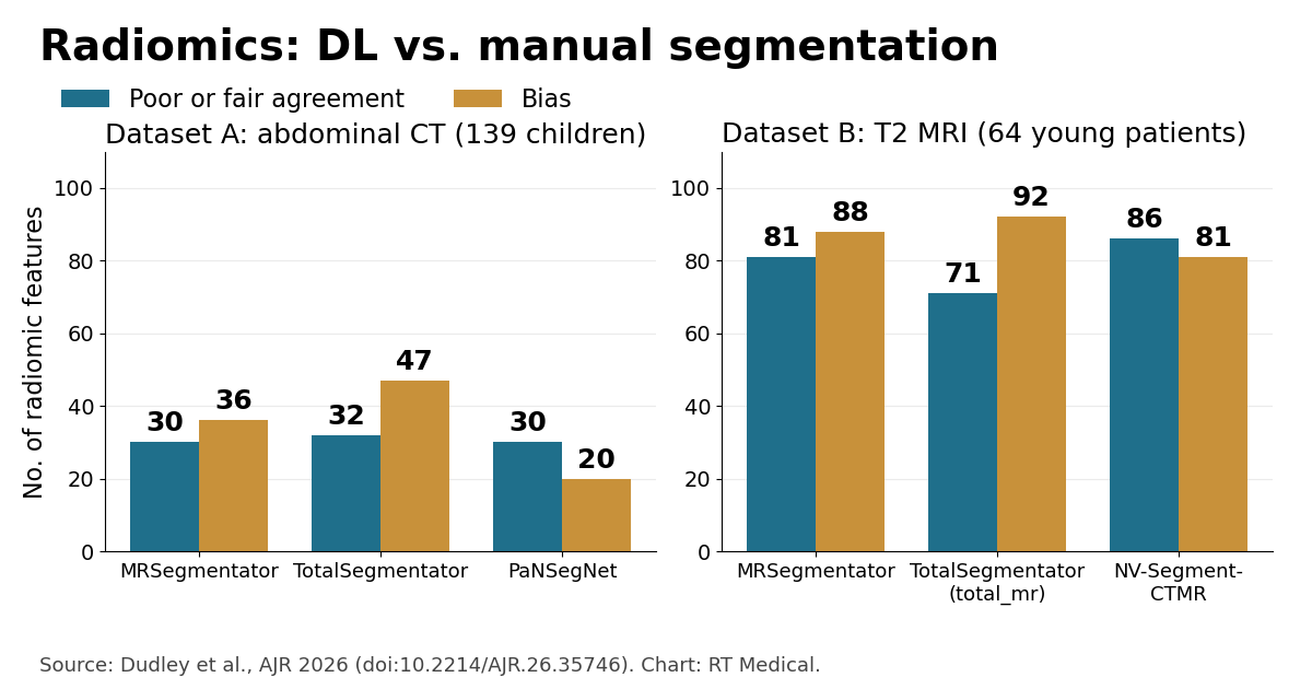
According to AuntMinnie’s coverage of the paper, the three models behaved similarly within each dataset. On pediatric CT (dataset A), 30 features with MRSegmentator, 32 with TotalSegmentator and 30 with PaNSegNet showed poor or fair agreement with the manual reference; 36, 47 and 20 features, respectively, showed bias. On MRI (dataset B) the picture was more pronounced: 81, 71 and 86 features with poor or fair agreement for MRSegmentator, TotalSegmentator and NV-Segment-CTMR, and 88, 92 and 81 with bias.
When those features fed predictive models, switching the segmentation source came at a cost. Compared with models built on manual contours, accuracy dropped by a mean of 0.05 with MRSegmentator and 0.12 with PaNSegNet; there was no significant difference with TotalSegmentator (mean, −0.01). Generalizability was worse in all three cases, with mean differences of 0.15 (MRSegmentator), 0.10 (TotalSegmentator) and 0.17 (PaNSegNet). The material available does not specify the exact scale of these metrics, so the values should be read as relative differences reported by the authors.
The group concluded that features from deep learning masks commonly show limited agreement with, and bias relative to, manual ones. They recommend that developers of radiomic models clearly describe their segmentation methodology, and that anyone validating or comparing models recognize that segmentation differences can confound results.
Technical background: why the mask boundary matters so much
This section is general domain knowledge, not study findings. Radiomics turns a region of interest into hundreds of numbers. Shape features (volume, surface area, sphericity) depend directly on the contour. First-order features (mean, standard deviation, skewness, histogram entropy) shift when a mask includes a few voxels of fat, vessel or adjacent parenchyma. Texture features, such as those derived from the gray-level co-occurrence matrix (GLCM) and from run-length and size-zone matrices, are particularly sensitive because the boundary concentrates the strongest intensity gradients in the image.
That is why two contours with a high Dice coefficient can still yield quite different texture features: Dice measures volumetric overlap but barely registers a thin rim of voxels at the periphery, exactly where much of the texture information lives. The Image Biomarker Standardisation Initiative (IBSI) standardized feature definitions and computation (Zwanenburg et al., Radiology 2020), which reduces variability across software packages but does not address variability introduced by the segmentation itself. Recent stories on this blog, such as the model that reads cancer risk in CT scans called normal and the one that predicts immunotherapy pneumonitis from CT, rely on precisely this segmentation-feature-model chain.
Practical implications for radiology and radiation oncology
For research groups, the message is methodological: mixing manual and automated masks in the same study, or externally validating a radiomic model with segmentations from a different source, can introduce a systematic shift that is easily mistaken for a biological effect. Recording the segmentation tool, version and task becomes as important as recording the acquisition protocol.
In radiation oncology, where automated contouring of organs at risk is already routine in many departments, the study echoes a familiar medical physics point: geometric metrics such as Dice and Hausdorff distance are necessary but not sufficient to judge whether a contour is fit for a specific downstream use, whether a dose-volume histogram or an imaging biomarker. Evaluation should consider the end use and keep structure nomenclature consistent, a topic covered in our TG-263 guide. Note that the study did not assess contours for treatment planning or dosimetry; the extrapolation to radiotherapy is contextual.
In Brazil and across Latin America, where radiomic cohorts are often assembled from multiple centers using open tools, the recommendation to document and standardize segmentation is especially relevant, since between-hospital differences can add to between-method differences.
Limitations and next steps
This is a research letter with moderate sample sizes, pediatric and young adult populations and retrospective data from earlier studies. Manual segmentation was treated as the reference, although it has its own inter-observer variability. The models tested are public, general-purpose tools with no fine-tuning on the evaluated datasets, and the findings do not speak to other tools, commercial or otherwise. The material consulted also does not report the total number of features extracted or the exact definitions of the agreement and performance metrics. As with other recent radiomics models, external validation and transparent methodological reporting remain prerequisites for clinical use.
Source: AuntMinnie



