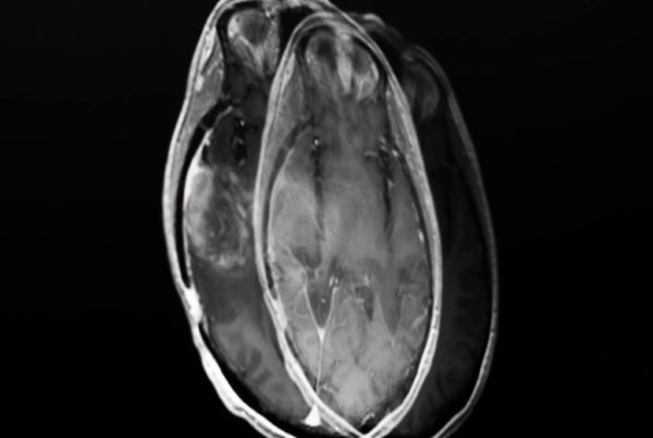
The window for thrombolysis in ischemic stroke has stopped being a clock and become an image. With updated American Heart Association and American Stroke Association guidelines extending clot-dissolving therapy from 4.5 hours to as long as 24 hours in patients selected by a favorable perfusion profile on CT, the limiting factor has moved: it is no longer time from symptom onset, it is whether the neurologist can actually see the images, wherever they happen to be. That bottleneck is what OSF HealthCare in Peoria, Illinois, is attacking with an AI-enabled enterprise imaging platform.
What changed in the guidelines
Arun V. Talkad, MD, ministry director of stroke at OSF HealthCare, laid out the logic in an interview with Diagnostic Imaging. The combination of updated AHA/ASA guidance with data from the OPTION trial, which showed benefit from CT perfusion-guided treatment out to 24 hours, lets facilities without in-house imaging expertise identify eligible patients well beyond the traditional window — provided they have connected software for sharing images.
It is worth unpacking what “favorable perfusion profile” means in practice, because that is where radiology enters. CT perfusion separates tissue already lost — the ischemic core, typically defined by relative cerebral blood flow below 30% — from tissue at risk but still salvageable, the penumbra, delimited by time to maximum of the residue function above 6 seconds. The candidate patient has a small core and a large penumbra: a favorable mismatch ratio says there is still a great deal to rescue. It is the same reasoning that underpinned the extended-window thrombectomy trials, now applied to thrombolytics.
The neurons-per-minute arithmetic
Talkad translates the urgency into numbers every on-call physician should carry around. “Every minute of stroke, we lose 1.9 million brain cells, and so over the course of an hour, just one hour of stroke, we lose 120 million brain cells. That’s the same amount of brain cells we lose in normal 3.6 years of aging,” he said.
That arithmetic changes how you read the workflow. If each hour of delay equals nearly four years of brain aging, the fifteen minutes spent burning a CD, the forty-minute ambulance ride to transfer the patient to where a neurologist sits, and the time lost trying to open a perfusion study in a viewer that will not load stop being administrative inconvenience. They are a dose of harm.
The bottleneck is not the algorithm, it is image access
The most interesting point in the interview is almost anti-technological. Before the platform, Talkad said, stroke specialists covering outlying referring hospitals frequently had to make treatment recommendations without seeing the images — deciding by phone, from someone else’s description. The platform’s core value, in his framing, is giving the remote physician the same visual access an on-site clinician would have.
In other words: the gain comes not from the algorithm deciding, but from the algorithm post-processing fast and delivering the result in a viewer that opens anywhere. That is the difference between AI as oracle and AI as plumbing — and the second is, in practice, far more transformative. The same logic showed up when we covered Epic’s feature letting hospitals share images without CDs: removing friction from data transport is worth more than any new accuracy metric.
Technically, this depends on distinctly unglamorous things. Server-side rendering, so the physician’s device receives processed images instead of downloading hundreds of megabytes of a perfusion study. A zero-footprint viewer with browser access. Push notification that opens the case, not an email saying a case exists. DICOMweb interoperability so each hospital’s PACS talks to the platform without bespoke integration.
From 25% to 80%: the size of the potential gain
Talkad’s projection is the strongest part of the argument. “Right now, only about 25 percent of patients show up within that first three- to 4.5-hour window that even allows us to be able to treat them, and if about 80 percent of people are showing up within 24 hours, now I might be able to treat 80 percent of patients. Obviously, I’m not going to treat all of them, but just tripling the amount of people that I could treat is going to now offer the benefit of having so many more people continue to go home.”
Read that number carefully: 80% is the population eligible for assessment, not the population treated. Perfusion selection then keeps those with favorable mismatch, no hemorrhage and no contraindication to thrombolysis. Talkad makes that caveat himself. Even so, moving from a 25% ceiling to an 80% ceiling at the triage stage is an order-of-magnitude change in the denominator — and that is what justifies expanding the platform from the initial three hospitals to all 18 in the system.
The equity dimension
Talkad stresses significant health equity implications, and the mechanism is direct. Rural and small hospitals have a CT scanner but rarely a neuroradiologist or vascular neurologist on call. Without a platform, a patient arriving at one of those sites has two bad options: go untreated, or be transferred, burning exactly the time that would define eligibility. With remote access to the processed images, the decision is made where the patient already is.
That is the same economic argument behind reimbursing triage tools — we followed the case when Medicare approved an add-on payment for CT triage AI. Once a health system recognizes that the value lies in shortening time to decision, the investment math closes.
Local reality and limitations
In Brazil, the national stroke care line and dedicated stroke units concentrate thrombolysis in accredited centers, and CT scanners are reasonably distributed. CFM Resolution 2,314/2022 provides the regulatory basis for telediagnosis and teleradiology. What is generally missing is neither regulation nor hardware — it is perfusion software deployed outside major centers, and enough bandwidth for the study to leave the hospital in minutes. A service still exporting DICOM to physical media captures none of the benefit of a 24-hour window.
The limits of the report also need stating. This is an interview with a clinical leader, not a comparative study: there is no published 90-day mRS outcome, symptomatic hemorrhage rate, or door-to-needle time before and after deployment at the three hospitals. Extending the window also widens the risk of selecting badly — thrombolysing a large-core patient late increases hemorrhage without functional gain, and the quality of perfusion post-processing becomes a direct patient-safety variable. Different software packages produce different core volumes from the same acquisition, a known standardization problem still unresolved.
The natural next step is publishing real deployment data: how many additional patients were treated, with what outcome and at what cost per case. Until that arrives, what exists is a solid architectural thesis — acute stroke treatment has become an imaging logistics problem — and an 18-hospital system betting on it.