

A Viewpoint article in JAMA Pediatrics makes a concrete proposal for anyone who follows children after traumatic brain injury (TBI): keep CT for the initial diagnosis, then move surveillance imaging to rapid MRI protocols that run in five to seven minutes without sedation. The case is dosimetric, not cosmetic. The head is the most frequently imaged body region in pediatric CT, and the evidence on cumulative cancer risk has become hard to wave away.
What the proposed imaging pathway changes
The authors lay out a three-branch clinical pathway, and each branch deserves a close look.
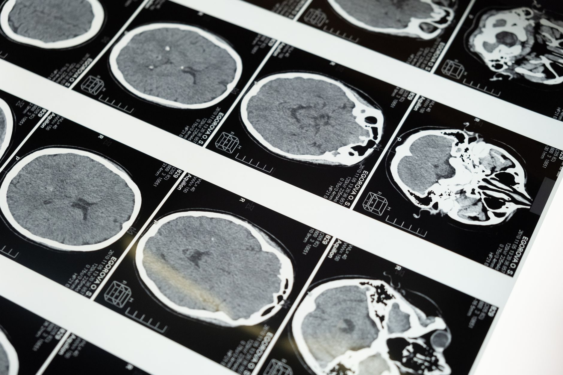
First, initial diagnosis still relies on index cranial CT. Nobody is proposing to retire the scanner at the emergency department door: CT is fast, available around the clock, and better for detecting acute hemorrhage and fracture. Second, stable patients who need repeat imaging should be routed to MRI rather than to a second or third CT. Third, for unstable patients the priority is timely imaging — whatever modality is at hand — plus urgent neurosurgical consultation. Protocol purity has no place in that scenario.
Sequence selection is the technical core. The authors recommend motion-tolerant, blood-sensitive sequences: T2 with gradient echo, or susceptibility-weighted imaging (SWI). The logic runs two ways. Short sequences reduce the need for sedation, itself a real risk in children, and SWI is notably more sensitive than CT for microhemorrhage and diffuse axonal injury — findings CT simply does not resolve.
Why cumulative dose moved to the center of the argument
What pushed this discussion onto the agenda was a retrospective cohort study published in the New England Journal of Medicine in September 2025. Investigators reconstructed the full imaging histories of roughly 3.7 million children born between 1996 and 2016 across six US health systems and Ontario, Canada, following them until cancer diagnosis, death, loss of coverage, or age 21.
The numbers are uncomfortable. One or two head CT exams were associated with a 1.8-fold higher risk of a hematologic malignancy diagnosis; with more exams — and therefore more dose — the ratio climbed to 3.5. Head CT, the most frequently performed CT in this age group, delivered a mean bone marrow dose of 13.3 mGy, with a relative risk of 1.35 (95% CI 1.23–1.48) and an attributable risk of 25.9%. At population level, the authors estimate that roughly one in ten pediatric hematologic malignancies may be attributable to radiation from medical imaging.
These findings deserve the caution any observational design demands: indication bias, estimated rather than measured dosimetry, and the real possibility that a scanned child differed from an unscanned one to begin with. Even so, few imaging specialists would argue with the practical conclusion — in pediatrics, CT dose is justified case by case, never by habit. That is ALARA taken seriously, in the spirit of the Image Gently campaign since 2008. Worth remembering that US CT radiation dose fell 22% over a decade, proof that optimization works once you measure it.
The barriers the proposal openly admits
This is not a naive article. The authors themselves list the obstacles: availability of MRI scanners and trained technologists, particularly outside daytime hours. A department that staffs CT overnight but runs MRI only on business hours cannot realistically offer follow-up MRI at 3 a.m. on a Saturday.
More importantly, the authors explicitly do not recommend transferring children to another facility solely to obtain an MRI. The risk of transporting a neurologically injured patient outweighs the benefit of avoiding radiation. The recommendation is conditional by design: where MRI is available, every effort should be made to use it; where it is not, well-optimized CT remains the right answer.
What it means in day-to-day practice
Outside major academic centers the bottleneck is familiar and lopsided: CT scanners are reasonably distributed, MRI far less so, and public-system access usually means a waiting list. That does not invalidate the pathway — it defines where it applies today. Pediatric referral hospitals and private services running MRI on extended hours can implement a rapid-MRI surveillance protocol with the equipment already on the floor.
On the radiology side, three moves unlock the process. Creating and clearly naming a dedicated protocol in the PACS (“pediatric TBI rapid MRI”) keeps the order from defaulting to a 30-minute full brain study. Training technologists in feed-and-wrap for infants and in awake-child handling sharply cuts sedation demand. And standardizing the report with fixed descriptors for hemorrhage, contusion, and diffuse axonal injury makes serial comparison far more reliable — the same reasoning behind our coverage of radiologist variability in reading lung CT.
There is a safety dimension too. Swapping CT for MRI does not remove risk; it swaps one risk for another. The magnetic field zone comes with its full burden of ferromagnetic screening. Our reporting on pediatric MRI accidents tied to protocol lapses shows that scaling pediatric MRI volume without reinforcing metal screening is a bad trade.
Outlook and limitations
The JAMA Pediatrics piece is a Viewpoint, not a formal guideline with graded evidence, and should be read that way: a proposed clinical pathway built on solid epidemiology and on earlier feasibility work with rapid MRI in pediatric head trauma. What is still missing is a pragmatic trial comparing hard outcomes — neurosurgical reintervention, functional sequelae, length of stay — between CT-based and MRI-based surveillance. Until that exists, the decision stays individualized.
The underlying trend is unmistakable, though. Pediatric radiology spent twenty years learning to cut dose per exam; the next gain comes from cutting the number of ionizing exams, replacing them wherever an equivalent alternative exists. For the most vulnerable patients of all, that math adds up.
Source: The Imaging Wire — CT vs. MRI for Pediatric TBI Surveillance



