

Eight in ten are comfortable with an AI risk score
A survey of 319 patients published Aug. 8 in Clinical Imaging produced a result that cuts against the expectation of resistance: 80.5% of women said they would be comfortable with AI-based breast cancer risk assessment, versus only 31.0% who preferred an estimate based on personal and family history. The research was led by Liliana Light, an MD student at Howard University in Washington, D.C. The finding matters because barriers to adopting these models are usually attributed to patient distrust — and distrust, here, did not show up.
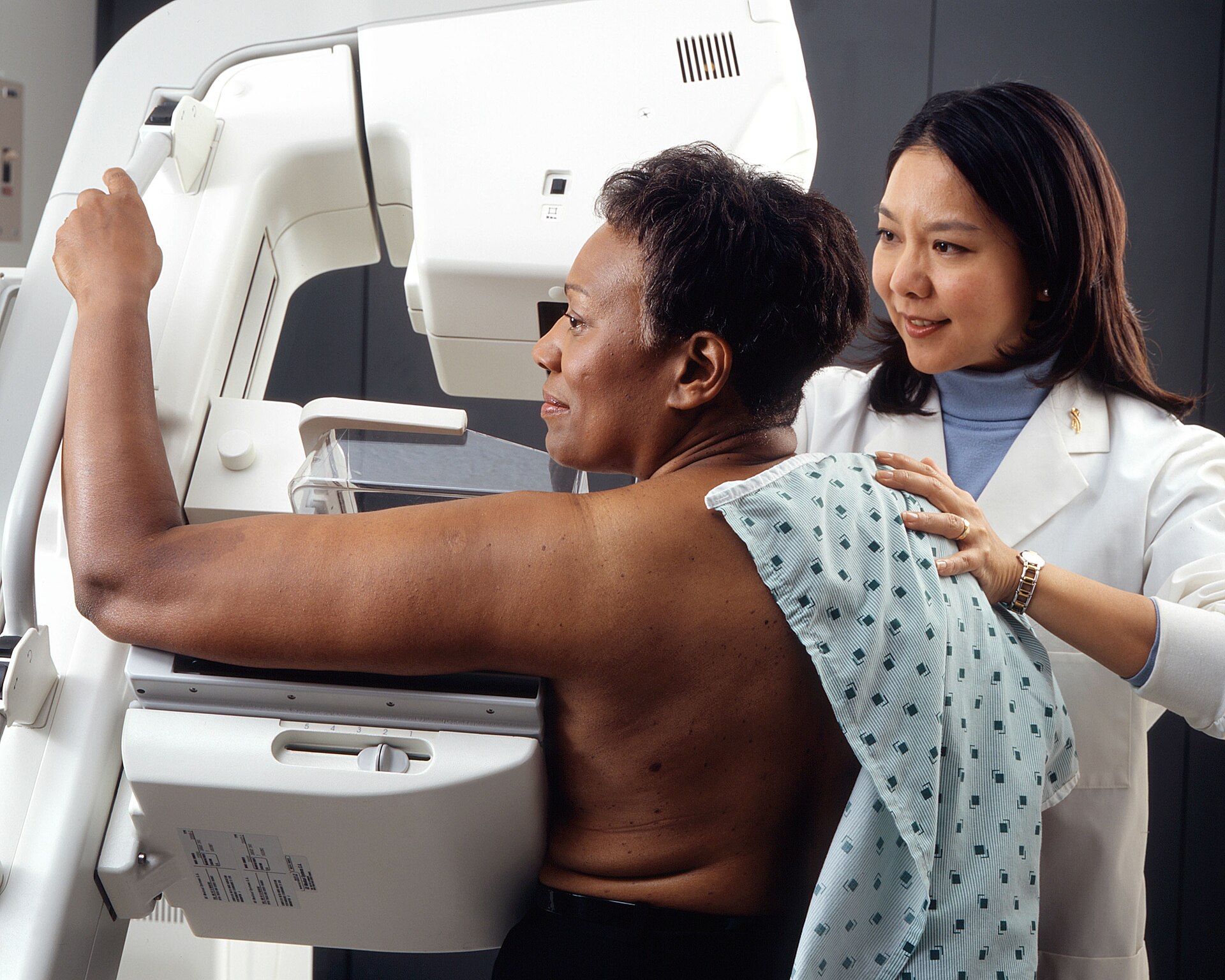
How the survey was conducted
The study collected 319 completed questionnaires from patients at four medical centers, spread across radiology, gynecology and internal medicine clinics. That distribution by clinic type is not an administrative detail — it produces the study’s most actionable finding, as we will see, because preferences shifted depending on where the patient was approached.
The design is cross-sectional and declarative: it measures stated comfort and intent, not observed behavior. The gap between those is worth recording. Telling a researcher you would accept an algorithmic risk estimate is different from receiving a score indicating elevated risk and deciding, with a full calendar and a copay to pay, to undergo supplemental MRI. Even so, measuring stated acceptance has immediate practical value: it is the number services use to decide whether implementing the workflow is worth it.
Who patients want delivering the result
The most useful part of the study is about communication, not technology. Preferences on who should convey the risk varied by recruitment clinic: among women approached in gynecology and radiology offices, 82.5% preferred receiving the result from their gynecologist and 57.3% from their radiologist. Among those recruited in the internal medicine clinic, most (63.3%) preferred their own primary care physician.
The reading is direct and slightly uncomfortable for radiology: patients want to hear the result from someone they already know. It is not a rejection of the radiologist — it is a preference for continuity. That has an immediate operational consequence, because the risk score is generated inside the imaging service, from the mammogram, while the conversation that gives it meaning has to happen in another office, with another clinician, who often does not know how the number was produced.
Any serious risk-AI deployment therefore carries a physician-to-physician communication component that project budgets tend to underestimate. Without a channel to explain to the gynecologist and the internist what that score means, the predictable outcome is a number circulating in the chart without driving management — or driving disproportionate management.
What women do when risk is elevated
Among women identified as being at increased risk, 90.3% expressed interest in additional screening tests. Interest in genetic testing also split by risk stratum: 72% among those at increased risk, versus 50% among those who were not.
Ninety percent stated uptake of supplemental screening is a number that demands capacity planning before deployment. If a service starts computing AI risk for everyone who has a mammogram, and a fraction of that population is classified as high risk, and nine of every ten of those women want an additional exam, demand for breast MRI and targeted ultrasound grows predictably. Again, the bottleneck is not acceptance — it is the schedule.
Interest in genetic testing among half of the women not at increased risk also deserves attention. It signals a demand for information that can outrun clinical indication, with implications for cost, genetic counseling and anxiety. No service should switch on a risk model without first deciding what it will do with patients who want more than the evidence recommends.
Technical context: what AI breast risk means
Distinguishing tool generations is worthwhile, because the difference explains the current interest. Classic models such as Gail and Tyrer-Cuzick estimate risk from clinical variables: age, menarche, parity, family history, prior biopsies, sometimes categorized breast density. They are questionnaire models — performance depends on history-taking quality and they notoriously underestimate risk in populations underrepresented in the derivation cohorts.
Image-based models work differently. A convolutional neural network processes mammographic pixels directly and learns texture patterns, density distribution and parenchymal architecture associated with future cancer diagnosis — including patterns that map to no descriptive category radiologists use. The gain is that the estimate does not depend on the patient knowing her aunts’ and grandmothers’ health histories, which matters especially in populations with incomplete family records.
The honest counterpoint is that these models inherit the bias of their training data. An algorithm derived mostly from white women in one health system has no guaranteed calibration in another population — and calibration, not discrimination, is what matters when the number will decide whether someone gets annual MRI. That this survey came out of a historically Black university with a diverse population makes the acceptance finding more interesting still: high acceptance in a group with historical reasons for distrust.
Practical implications for imaging services
Three consequences emerge for anyone considering deployment. First, define the communication path before the software. Because patients prefer hearing results from the gynecologist or internist, the report must deliver the score in a form interpretable by non-radiologists — with a risk band, comparison against population average and suggested management, not just a bare number.
Second, size the induced demand. Simulate how many additional MRIs and ultrasounds the model would generate when applied to your actual population, and compare with installed capacity. Doing that math beforehand avoids the classic situation of identifying risk and being unable to offer the exam that identification recommends.
Third, align expected benefit. It is worth remembering what happened with detection AI: a recent survey showed that mammography AI delivered less than radiologists expected on recall, biopsy and burnout. Risk models are a different and better-positioned promise — stratifying who needs more, rather than re-reading what was already read — but they deserve the same methodological skepticism in evaluation.
Limits and next steps
The limitations are typical of surveys: convenience sample at four centers, self-selection bias among those willing to answer, and the fact that stated comfort does not predict actual uptake. Health literacy effects were not measured, nor was question framing tested — asking whether someone “would feel comfortable” with AI tends to produce higher rates than asking whether she “would trust AI more than her physician.”
The resulting research agenda is clear: measure observed rather than stated uptake; assess risk model calibration in diverse populations; and test risk communication formats that work for the non-radiologist clinician. What this study establishes, usefully, is that “will the patient accept it?” can come off the list of obstacles. She accepts. What remains unanswered is whether the health system is ready for what she will ask for next — as unanswered as which supplemental modality actually adds value in screening or how to fold in newly cleared tools such as DeepHealth’s AI for breast ultrasound.
Source: AuntMinnie



