

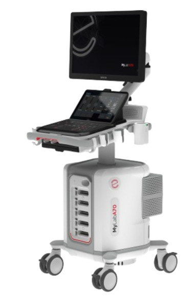
Modern premium ultrasound has evolved from a probe that simply produces pictures into a full clinical decision platform: today’s high-end systems fuse on-board artificial intelligence, automated measurements, shear wave elastography and contrast-enhanced ultrasound (CEUS) to answer, quantitatively, questions that once demanded MRI, CT or a biopsy. Systems such as Esaote’s MyLab A70 are a concrete example of this generation, but what matters most for the radiologist is the physics and the workflow behind the acronyms.
On-board AI: the ultrasound that measures on its own
The short answer to “what does AI do in ultrasound” is: it removes variability. AI-assisted automation such as Esaote’s Augmented Insight recognizes anatomy, places calipers and computes standardized measurements without relying solely on the operator’s hand. In obstetrics, algorithms identify the correct fetal planes and measure head circumference, biparietal diameter and femur length automatically; in cardiology, they estimate ejection fraction from an automatic left-ventricle trace; in thyroid and breast work, they score nodules against structured criteria.
The payoff is not only speed. Because the measurement no longer hinges on individual judgment, two studies acquired by different sonographers tend to converge, which is decisive when tracking a lesion over time. That same drive toward standardization fuels the heavy investment in AI for imaging now reshaping the market and places sonography inside the broader digital transformation of radiology that already reorganized PACS and structured reporting.
Shear wave elastography: quantifying tissue stiffness
Elastography answers a blunt clinical question: how stiff is this tissue? Fibrotic or malignant tissue is usually harder than healthy tissue, and stiffness has become a non-invasive biomarker. In shear wave elastography the transducer fires an acoustic push pulse (acoustic radiation force) that displaces tissue by a few micrometers. That displacement launches shear waves travelling laterally, and the scanner tracks their speed using ultrafast ultrasound pulses.
Shear wave speed (c) is converted into stiffness through the Young’s modulus, roughly $E = 3\rho c^2$, where $\rho$ is tissue density. Results are reported in kilopascals (kPa) or in m/s and displayed as a color map overlaid on the grayscale image. Modern systems also flag measurement reliability, discarding regions where the shear wave propagates inconsistently so the clinician trusts only well-sampled values. The most established application is staging liver fibrosis, where elastography now replaces many biopsies in chronic liver disease and enables painless follow-up; it also helps characterize breast and thyroid nodules and supports musculoskeletal assessment.
Contrast-enhanced ultrasound (CEUS): perfusion in real time
CEUS adds the vascular dimension. The contrast agent is made of gas-filled microbubbles a few micrometers across, smaller than a red blood cell, wrapped in a phospholipid shell; injected intravenously, they stay within the vascular compartment. When the ultrasound beam hits them at a low mechanical index, the microbubbles oscillate non-linearly and return harmonic signals that the scanner separates from surrounding tissue, revealing blood flow down to the microcirculation.
Unlike CT iodinated contrast or MRI gadolinium, ultrasound contrast is cleared through respiration and is not nephrotoxic, making it safe in patients with renal impairment. Clinically, CEUS characterizes focal liver lesions by their real-time enhancement pattern, assesses the kidneys, checks vascular graft patency and helps distinguish tumor from thrombus. Because it happens at the bedside and in real time, it adds functional information without moving the patient to another suite.
Ergonomics, workflow and clinical breadth
A premium system is also defined by ergonomics. Adjustable consoles, touchscreens, customizable protocols and a broad transducer portfolio let one machine serve general radiology, women’s health, cardiology, vascular and musculoskeletal imaging. Reducing repetitive motion matters: repetitive strain injuries are common among sonographers, and an ergonomic design protects the person operating the machine dozens of times a day.
Workflow extends beyond the scanning room too: DICOM connectivity, PACS integration and automated transfer of measurements into the structured report turn the exam into reusable data. In that ecosystem it pays to choose FDA-cleared AI vendors, whose algorithms passed regulatory validation before they influence a clinical decision.
Access, cost and the future of point-of-care automation
Ultrasound is the most accessible and widely distributed imaging modality worldwide, from tertiary hospitals to primary-care clinics. Features once reserved for flagship consoles, such as elastography and AI automation, are migrating into mid-range and portable units, widening access to quantitative diagnosis outside major centers. AI-assisted automation in point-of-care ultrasound (POCUS) is especially promising: by guiding acquisition and confirming that the imaging plane is correct, it flattens the learning curve so that non-expert clinicians can capture reliable diagnostic images.
The trajectory is clear: less operator dependence, more quantification and tighter integration with the rest of the imaging journey. Platforms like the MyLab A70 illustrate the direction, but the real value lies in understanding the physics of elastography, the mechanism of microbubbles and the role of AI, so each feature is used for the right indication, always with clinical validation and quality control.
Source: DOTmed News


