

When AI Solves a Problem Physics Alone Could Not
A consortium of researchers from the University of Rochester, Brown University and the University of Copenhagen has published in Science Advances a technique that could change how we diagnose neurodegenerative diseases. Named MR-AIV (Magnetic Resonance Artificial Intelligence Velocimetry), it uses physics-informed AI to reconstruct, voxel by voxel, the three-dimensional velocity field of brain fluid from dynamic contrast-enhanced MRI. It is the first non-invasive method capable of mapping the human glymphatic system without direct velocity measurements.
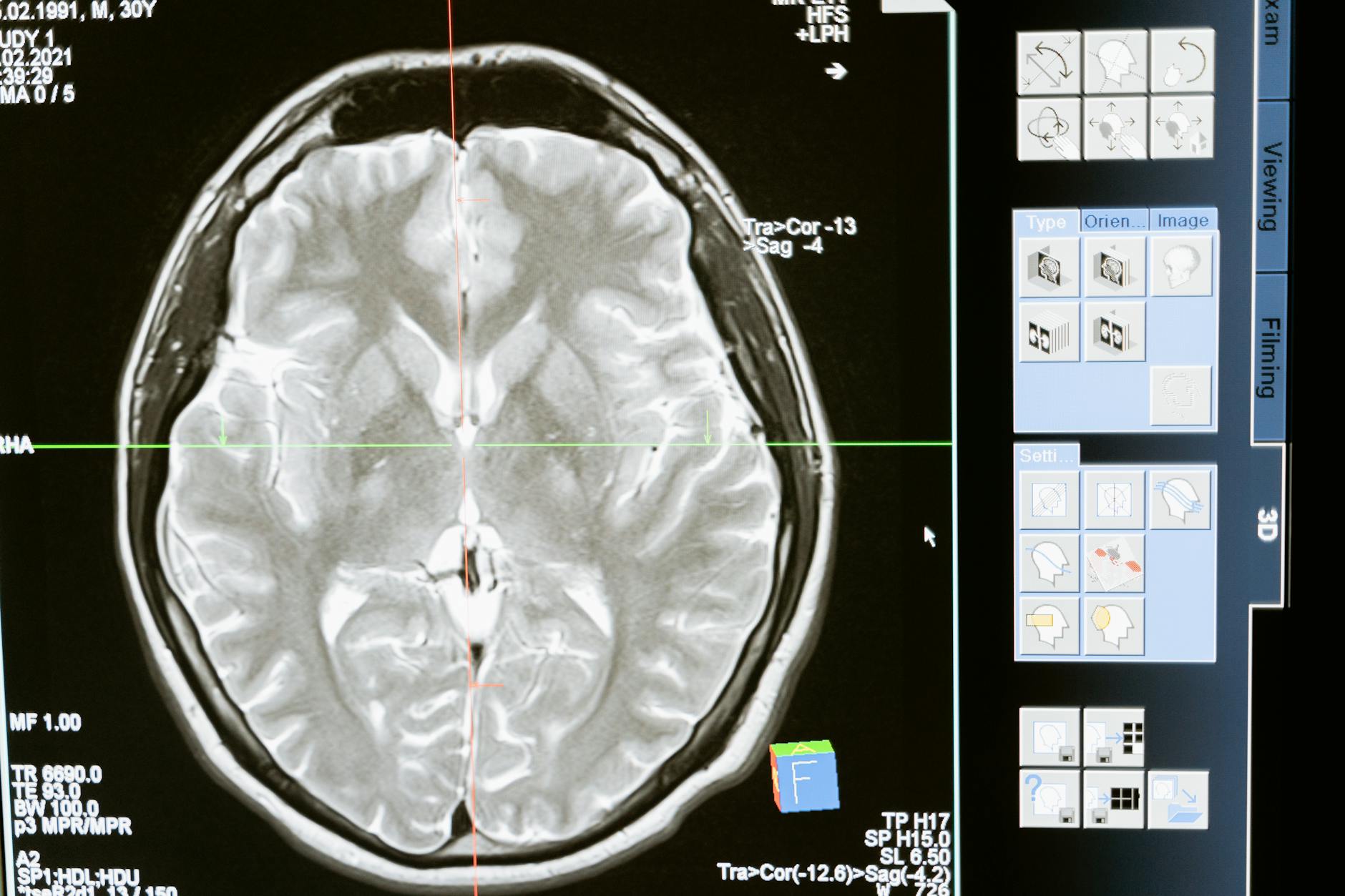
The team is led by Douglas Kelley (Department of Mechanical Engineering, Rochester), Juan Diego Toscano (Brown PhD student) and George Karniadakis (Brown professor). The paper, published with DOI 10.1126/sciadv.aeb0404, has direct relevance for radiology: the technique runs on existing clinical equipment and gadobutrol, a widely used paramagnetic contrast agent.
How MR-AIV Works
Glymphatic flow — the system that flushes the brain of waste proteins, including the amyloid-beta linked to Alzheimer’s — operates at extremely low velocities, below the detection threshold of conventional MR techniques such as phase contrast. MR-AIV attacks the problem indirectly: it observes the time evolution of gadobutrol concentration after intrathecal or intravenous injection and uses a physics-informed neural network to infer the velocity, pressure and permeability fields that best explain the observed diffusion.
The trick lies in the physical constraints baked into the network. Rather than learning purely statistical patterns, the model is forced to obey the Navier–Stokes equations and the mass transport equation. That sharply reduces the solution space and lets the algorithm extract velocity where the image alone shows no movement. It is a canonical application of physics-informed neural networks (PINNs) — a field where Karniadakis is a global reference.
What the Glymphatic System Reveals
Results show that the glymphatic system operates at two distinct speeds. There is a fast track, with average velocity around 3 µm/s, running through the superficial regions between the skull and the brain. And there is a slow track, around 0.1 µm/s — roughly 50 times slower — through deep brain tissue. That velocity contrast was previously inaccessible to any non-invasive imaging technique.
The clinical relevance is direct. During deep sleep, the glymphatic system ramps up and clears toxic proteins such as amyloid-beta and tau. Dysfunction in this system is linked to Alzheimer’s, Parkinson’s, ALS and to cognitive impact from traumatic brain injury. “We hope to someday be able to see whether an Alzheimer’s patient has poor circulation in their brain, or screen for poor circulation earlier in life to try to stave off Alzheimer’s,” Kelley said.
What Changes for Clinical Radiology
Three practical implications stand out. First, MR-AIV does not require proprietary hardware — it can run on installed 3T MRI scanners with adapted DCE sequences. That brings the technique close to the real clinical world, unlike solutions requiring new gradient systems or dedicated coils. Second, it opens a window for early biomarkers of neurodegeneration, complementing amyloid PET and plasma tau biomarkers. Third, it positions physics-informed AI as a diagnostic tool, not merely a productivity amplifier.
For radiologists tracking AI adoption, the study dialogues with discussions we explored in our piece on whole-body AI MRI predicting disease years ahead and with the analysis of the five strategic questions for AI adoption in radiology. The common thread is clear: the frontier of imaging has shifted from pure spatial resolution to inferring invisible physiological processes.
Limitations and Next Steps
The study has candid limitations. Current human validation uses a small number of volunteers, DCE-MRI acquisition is still time consuming, and the use of gadolinium for this protocol must be clinically justified case by case. There is also the regulatory discussion around intrathecal contrast in prospective studies. The bridge from proof of concept to clinical protocol will require multicenter trials, comparisons with CSF biomarkers and amyloid PET, and definition of velocity cut-offs for different age brackets.
Another challenge is operational: MR-AIV depends on good signal-to-noise across sequential acquisitions, which lengthens scanner time per patient. In services with tight schedules, that becomes a cost variable. But in neuroradiology centers of reference, the potential diagnostic gain justifies the investment. The project’s next phase includes application in mild cognitive complaint patients and in chronic post-traumatic cases.
Outlook and Broader Impact
For radiology globally, three points matter. First, the installed base of 3T MRI in academic centers is compatible with DCE protocols — the bottleneck will be local development and validation of physics-informed AI pipelines, not hardware. Second, integration with plasma biomarkers already in use (p-tau217, GFAP) positions academic centers worldwide for interesting pre-clinical Alzheimer cohort studies. Third, the cost-effectiveness discussion must engage public and private payers early to avoid the technique becoming restricted to a high-cost private niche.
MR-AIV is, in essence, a reminder that radiology, computational physics and neuroscience are converging fast. Expect a wave of publications across 2026 and 2027 testing the approach in other pathologies — hydrocephalus, Parkinson’s disease and traumatic brain injury. Brain imaging just acquired a new temporal dimension, and it will change diagnostic criteria sooner than many anticipate.
Source: AuntMinnie — AI-powered MRI technique maps brain fluid flow tied to Alzheimer’s | Science Advances — MR-AIV



