

The problem: pancreas remains an oncologic blind spot
A new artificial intelligence model has outperformed radiologists at detecting early signs of pancreatic cancer on computed tomography (CT), according to a study covered by Health Imaging. The result reinforces one of the most promising fronts of AI applied to medical imaging: identifying small, pre-clinical lesions in one of the most lethal and silent malignancies in oncology.
Why this matters for radiology
Pancreatic ductal adenocarcinoma (PDAC) carries a difficult prognosis, with five-year survival historically below 12% in international statistics. Most cases are diagnosed at advanced stages, when metastases already preclude curative surgery. Only an estimated 15% to 20% of patients present with resectable disease at the time of diagnosis.
The compounding issue is that many cases show subtle clues on abdominal CT performed for other reasons — abdominal pain, acute pancreatitis, digestive complaints — months before the formal diagnosis. Findings such as pancreatic ductal dilation, focal parenchymal atrophy, contour changes or small isoattenuating hypodensities are easy to underestimate in high-volume routines.
What these AI models actually do
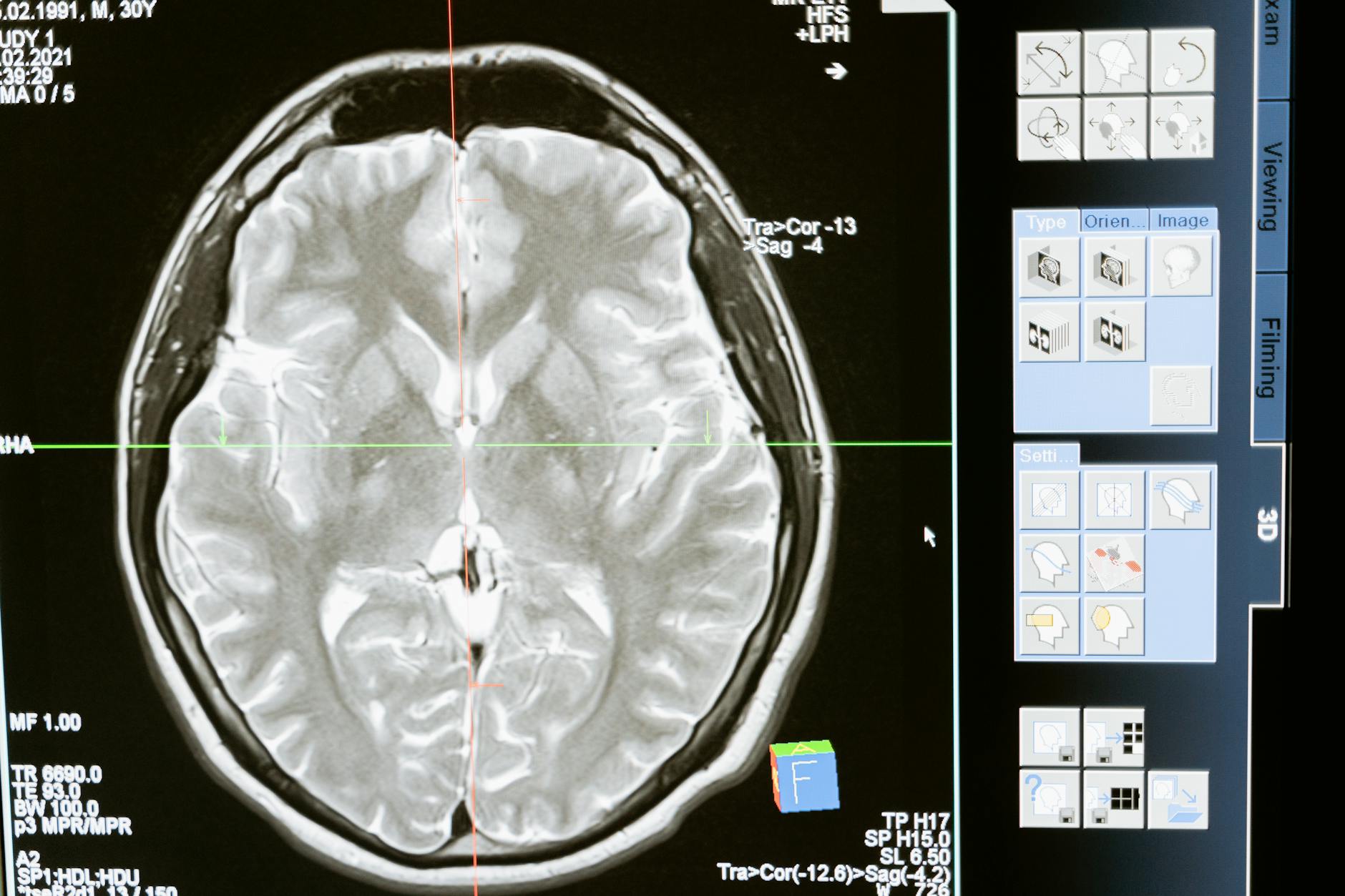
Recent AI models for pancreatic cancer detection follow three main lines. The first trains deep convolutional neural networks (CNNs) on contrast-enhanced CT in the pancreatic phase to identify focal lesions and distinguish them from healthy tissue. The second leverages segmentation architectures (U-Net and variants) to delineate parenchyma, pancreatic duct and potential lesions, turning the read into quantitative maps. The third combines both into multi-task pipelines: the algorithm segments the pancreas, measures features like volume, attenuation and ductal dilation and produces a suspicion score.
The differentiator is sensitivity to pre-clinical patterns: the model learns, from retrospective cohorts, how the gland evolves months before the clinical diagnosis. In studies published in recent years, this kind of pipeline has reached sensitivity close to or above that of experienced radiologists for tumors smaller than 2 cm — historically the hardest part of routine reads.
The link to opportunistic radiology
This advance fits into the broader trend of opportunistic radiology, in which exams ordered for other indications are repurposed for population-level screening. The same chest CT that catches a pulmonary nodule can flag coronary calcium and abnormal bone density; the abdominal CT ordered for diffuse pain can show ductal dilation or focal contour change. The discussion mirrors what appears in incidental findings on chest CT and in AI applied to plain radiography for clinical prediction.
The big promise here is to anticipate diagnoses by months or even years, with no extra cost to the system, by reanalyzing exams that already exist. For pancreatic cancer, where the curative window is narrow, gaining 6 to 12 months can change the natural history of the patient.
How to integrate this AI into the workflow
Bringing a pancreatic detection AI into routine practice requires some clear decisions. First, define which exams the algorithm runs on — typically contrast-enhanced abdominal CT in portal or pancreatic phase, with 1 to 3 mm slices. Second, choose how the result is delivered: a notification to the radiologist (“PDAC suspicion, review parenchyma and duct”), a pre-segmentation layer in the PACS or simply an annotation for auditing.
Third comes the conversation about false positives. High-sensitivity models can flag patients with chronic pancreatitis, benign cystic lesions or anatomical variants. Combining the score with clinical criteria — age, risk factors, lab abnormalities — cuts the noise and improves clinical adoption. The pattern is similar to what we have seen in established AI tooling in radiation oncology: real-world value only emerges when the model output is grafted onto clinical reasoning.
Limitations that still need to be addressed
Despite the optimism, the caveats are multiple. First, prospective multicenter validation remains scarce. Most studies are retrospective, with selected cohorts, which can overstate accuracy in real populations. Second, generalization across CT vendors and protocols is challenging: scanner, slice thickness, contrast type and acquisition phase all change parenchymal appearance.
There is also the regulatory side. Most published models are still research-grade, without clinical clearance for hospital deployment. And finally, integration with legacy systems — older PACS, RIS without orchestration capability, missing DICOM SR — remains a practical hurdle in many services.
What changes for the radiologist in the short term
Even before fully approved solutions arrive, a careful read of the pancreas on CTs ordered for other reasons becomes a topic of attention. Building a mental checklist — contour, focal atrophy, ductal dilation, subtle hypoattenuations, peripancreatic fat changes — captures part of the gain AI promises. Multidisciplinary discussion also gains weight: patients over 50 with new-onset diabetes, unexplained weight loss or acute pancreatitis without obvious cause deserve a careful review of pancreatic parenchyma.
In parallel, imaging managers can prepare by evaluating vendors that offer pancreatic detection modules, even if still research-grade. Having them mapped makes purchasing decisions easier when regulatory approval matures.
Outlook: early detection as a global priority
Pancreatic cancer has been climbing the research agenda, partly because therapeutic gains over recent decades have been modest. Detecting it earlier — when curative surgery is still possible — has become the bet with the highest population-level potential. AI applied to CT, alongside biomarkers and multimodal risk models, is one of the most promising fronts for shifting that statistic.
For the next few years, the agenda will be to integrate these technologies with structured secondary prevention, prospective trials and clinical governance. The first signs are on the table, and the result reported by Health Imaging is one more step in that direction.
Source: Health Imaging


